Karnataka 1st PUC Biology Question Bank Chapter 21 Neural Control and Coordination
1st PUC Biology Locomotion and Movement One Marks Questions and Answers
Question 1.
Expand CSF.
Answer:
Cerebro Spinal Fluid.
![]()
Question 2.
Name tie disorder characterised by the loss of motor function in human.
Answer:
Paralysis.
Question 3.
Name-the part of human brain concerned with homeostatic functions.
Answer:
Cerebellum
Question 4.
Which part of CNS is concerned with reflex action?
Answer:
AM, Medulla oblong and spinal cord.
![]()
Question 5.
Mention the arrungeetestt of grey matter and white matter in the spinal cord.
Answer:
In the spinal cord there will be inner grey matter and outer white matter.
1st PUC Biology Locomotion and Movement Two Marks Questions and Answers
Question 1.
Draw a neat labelled diagram of T.S. of spinal cord.
Answer:
Question 2.
Describe the structure of cerebellum.
Answer:
It consists of two lateral halves called cerebellar hemispheres. They are joined together by an elongated worm like median structure called vermis. The cortex, or the thin surface of the cerebellum consists of grey matter. Its surface is heavily folded and pleated and the folds are Called folia. It encloses white matter called cerebellar medulla which resemble a tree, called arbor vitae.
![]()
Question 3.
Compare the following:
(a) Central neural system (CNS) and Peripheral neural system (PNS).
(b) Resting potential and action potential.
Answer:
(a) Central Nervous System (CNS): This includes brain and the spinal cord.
(b) (i) Resting Membrane Potential:
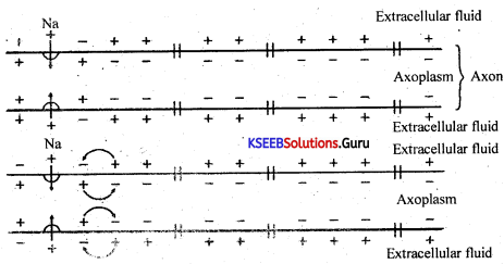
At the resting stage i.e., when the neuron is not involved in the conduction of impulses, the membrane of neuron is more permeable to K+ ions than to Na+ ions. The membrane is impermeable to the negatively charged proteins of the axoplasm and also Cl– ions. So the axoplasm (interior) of the axon has a high concentration of negatively charged proteins, Cl– ions and potassium ions but a low concentration of Na ions. Contrary to this, the extracellular fluid (exterior) has a high concentration of Na ions and low concentration of K+ ions.
These ionic gradients across the membrane, are maintained by the active transport of ions by the sodium-potassium pump, which transports three Na outwards and two K+ inwards. So the outer surface of the axonal membrane has a positive chárge and the inner surface of the membrane elaxoplasm is negatively charged the membrane is said to be polarised.
Action Potential:
When a thrishold stimulus is applied at a site on the polarised membrane, the membrane permeability at that site changes and becomes freely permeable to Na+ ions.
As a result, there is a rapid inflow of Na ions and there is a reversal in the polarity of the membrane.
The interior/axoplasm now becomes positively charged while the exterior becomes negatively charged and this is called depolarisation.
![]()
The potential difference across the membrane at the site of stimulation is known as action potential.
Now the current flows through the axoplasm from the depolarised region to the depolarised region.
As a result the action potential is generated in the next segment and this process continues along the length of the nerve fibre.
Question 4.
Differentiate between:
(a) Myelinated and non-myelinated axons.
(b) Dendrites and axons.
(c) Thalamus and Hypothalamus.
(d) Cerebrum and Cerebellum.
Answer:
(a) Thus there are a variety of differences between the myelinated and non- myelinated axons that impacts the transfer of nerve impulses.
(b) Most neurons have a cell body, an axon, and dendrites. The cell body contains the nucleus and cytoplasm. The axon extends from the cell body and often gives rise to many smaller branches before ending at nerve terminals. Dendrites extend from the neuron cell body and receive messages from other neurons.
(c) The thalamus also plays an important role in regulating states of sleep and wakefulness. … The hypothalamus controls body temperature, hunger, thirst, fatigue, sleep, and circadian cycles. The epithalamus functions as a connection between the limbic system and other parts of the brain.
(d) The cerebellum makes up the remaining part of the brain. The cerebrum controls voluntary movement, intelligence and memory.
![]()
Question 6.
Answer the following:
(a) Which part of the human brain is the most developed?
(b) Which part of our central neural system acts as a master clock?
Answer:
(a) Cerebrum
(b) Pineal gland.
Question 7.
Distinguish between:
(a) Afferent neurons and efferent neurons.
Answer:
(a) Afferent neurons carry signals to the brain and spinal cord as sensory data. … This neuron’s response is to send an impulse through the central nervous system. Efferent neurons are motor nerves. These are motor neurons carrying neural impulses away from the central nervous system and toward muscles to cause movement.
Question 8.
Write short notes on the following: [ Two marks each].
(a) Forebrain.
(b) Midbrain.
(c) Hindbrain.
(d) Synapse.
Answer:
(a) Fore Brain (Procencephalon): Includes cerebrum and diencephalon (Thalamus and Hypothalamus)
(b) Mid Brain (Mecencephalon): It is a highly reduced part in human.
(c) Hindbrain (Rhombencephalon): It includes cerebellum, ponsveroli and medulla oblongata (Stalk of the brain).
(d) A synapse is the functIonal junction between two neurons, formed by the membranes of a pre-synaptic neuron and a post-synaptic neuron.
1st PUC Biology Locomotion and Movement Three Marks Questions and Answers
Question 1.
Explain the following processes:
(a) Depolarisation of the membrane of a nerve fibre.
(b) Conduction of a nerve impulse along a nerve fibre.
(c) Transmission of a nerve impulse across a chemical synapse.
Answer:
(a) When an electrical stimulus is given to a nerve fibre, an action potential is generated. The membrane becomes permeable to sodium ions than to potassium ions. This results into positive charge inside and negative charge outside the nerve fibre. Hence, the membrane is said to be depolarized.
(b) Conduction of nerve impulse occurs due to the presence of active and electronic potentials along the conductors. Transmission of signals internally between the cells is achieved through a synapse. The ionic currents pass through the two cell membrane when the action potential reaches the stage of such synapse.
(c) At chemical synapses, impulses are transmitted by the release of neurotransmitters from the axon terminal of the presynaptic cell into the synaptic cleft. … Multiple cytosolic proteins including synapsin recruit synaptic vesicles to the active zone of the plasma membrane adjacent to the synaptic cleft.
![]()
1st PUC Biology Locomotion and Movement Five Marks Questions and Answers
Question 1.
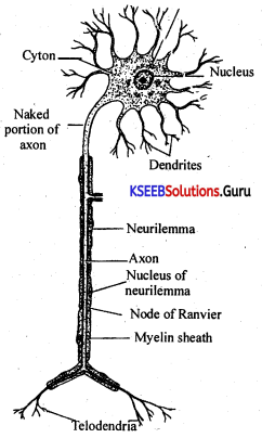
Explain the structure of a multipolar myelinated neuron.
Answer:
Neuron consists of two important regions.
(i) Cyton
(ii) Axon.
(i) Cyton : It is the cell body which is spherical or oval in shape. It contains a nucleus at the center and surrounded by cytoplasm. Cytoplasm is called Neuroplasm which contains minute fibers in a network and also special bodies called Nissl granules. These granules are believed to take part in protein synthesis. Periphery of the cyton produces branched processes. These short branches of cyton are called dendrites. One of these branches is much elongated and is called axon.
(ii) Axon : Axon is the elongated branch originating from a region called Axon hillock. The axon gives off branches at the end, called telodendria. Usually axon is covered by a continuous sheath called myelin sheath or medullary sheath. Schwann cells are present which are called neurilemma. This layer of Schwann Cells shows number of constrictions. These constrictions are called Nodes of Ranvier.
![]()
Question 2.
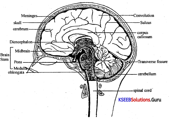
Draw a neatly labelled diagram of sagittal section of brain.
Answer:
Question 3.
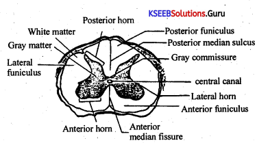
With a labelled diagram, describe the structure of human spinal cord.
Answer:
Tubular posterior extension of medulla oblongata is called the spinal cord. It comes out of the skull through an opening called ‘Formen magnum’ and is lodged within a canal of the vertebral column. As in brain, spinal cord is also covered by meninges such as piameter, archnoidmenter, durameter and CSF.
Cross section of spinal cord shows outer white matter and inner grey matter. White matter comprises of nerve fibres and grey matter comprises of cell bodies. Grey matter covers a central canal and appears in the form of letter H. In spinal cord there is a prominent cleft in the ventral side called ventral fissure. Similarly there is a thin separating line on the dorsal side called dorsal sulcus.
31 pairs of spinal nerves arise from the spinal cord. Each nerve is formed by the junction of two roots. The dorsal root is little swollen and comprises sensory nerve fibre and the ventral root consists of motor nerve fibres. Within this spinal cord the dorsal and ventral roots are connected by adjustor neurons.
Question 4.
Describe briefly the structure of human brain.
Answer:
Human brain is a soft nervous structure, weighing about 1280 to 1380 gms. It is protected by cranium of the skull and meninges. Meninges are the three membranes covering central nervous system (brain and spinal cord).
Human brain comprises of about 1000 billion neurons and is divisible into,
1. Fore brain (Prosencephalon).
2. Mid brain (Mesencephalon).
3. Hind brain (Rhombencephalon).
Sagittal section of brain reveals the following details.
Fore Brain : Fore brain comprises of cerebrum, diencephalon, (thalamus and hypothalamus) and olfactory lobes.
Cerebrum : It is the largest part of the brain. It consists of two hemispheres. Cerebral cortex is chiefly composed of cell bodies (cyton) of the neurons and is called grey matter, whereas cerebral medulla is chiefly composed of nerve fibres and is the seat of intelligence. Surface of the cerebral cortex shows number of folds and deep grooves . Folds of the cerebral cortex are called ‘gyri’ and the deep grooves are called ‘fissures’. Cerebrum also shows shallow furrows termed as ‘sulci’. These fissures divide each cerebral hemisphere into five lobes, namely.
![]()
(i) Frontal lobe : It is the largest of the four lobes making up the anterior one third of each hemisphere.
(ii) Parietal lobe : It lies behind the frontal lobe and is separated from it, by a fissure of Rolando or central fissure.
(iii) Temporal lobe : It lies below and behind the frontal lobe and is seperated from it by a lateral sulcus.
(iv) Occipital lobe : It lies behind the parieto-occipital sulcus.
(v) Insula (Island of Reil) : It is not visible because it is located deep with in the lateral sulcus. It is covered by parts of frontal, parietal and temporal lobes.
Corpus callosum : It is the network of nerve fibres connecting two hemispheres of cerebrum.
Diencephalon : Diencephalon is the part of a fore brain which connects the cerebrum with mid brain. Diencephalon comprises of thalamus and hypothalamus.
(i) Thalamus : It is the region of diencephalon which receives all the sensory stimuli and relays them to different areas of the cerebral cortex.
(ii) Hypothalamus : It is the region of the diencephalon found below the thalamus. It is associated with pituitary and pineal glands (pineal gland secretes the hormone melatonin). A stalk like infundibulum connects it with the pituitary gland, it controls various metabolic activities like regulation of the heart beat, regulation of body temperature, regulation of sleep, B.P, water electrolyte balance, etc.
Optic chiasma : Just below the hypothalamus there is a region at which the optic nerves cross each other. This region is called optic chiasma.
Olfactory lobes : These are extended part of the frontal lobes. They are olfaction in function and are poorly developed in human beings.
![]()
Mid Brain : It comprises of cerebral peduncles and corpora quadrigemina. Cerebral peduncles connect brain stem (midbrain, pons and medulla oblongata) with the cerebrum, where as corpora quadrigemina extend as optic lobes. These are centers for visual and auditory reflexes.
Hind Brain : Hind brain comprises of cerebellum, pons varoli and medulla oblongata.
Cerebellum : It is the first part of hind brain, and also the second largest part of the brain. It has two lobes. Cerebellum is concerned with co-ordination and smoothness of movement. It helps to maintain body balance and co-ordinated speech.
Medulla oblongata : It is the last part of the hind brain which posteriorly continues as spinal cord. It controls respiratory activities, dilation of blood vessels (It initiates impulses that cause constriction and dilation of the blood vessels), reflexes such as sneezing, coughing, vomitting and winking. Medulla oblongata also has control over heart beat and secretory activities of the alimentary canal.
Pons varoli: It is the bundle of nerve fibres situated between midbrain and medulla oblongata. It also bridges two lobes of the cerebellum. It regulates respiration.
Different parts of the brain contain inner spaces. These inner spaces of the brain are called ventricles. They are filled with Cerebro Spinal Fluid (CSF). The first and second ventricles are in the hemispheres of the cerebrum. Third one in the diencephalon and fourth in the medulla oblongata.
![]()
Question 5.
Draw labelled diagrams of the following:
(a) Neuron
(b) Brain.
Answer:
(a)
- Neurons or nerve cells are the functional units of the nervous system.
- Groups of neurons in the central nervous system are called nuclei.
- Groups of neurons in the peripheral nervous system are called ganglia.
- Multi polar nerve cells have many short dendrites and one long axon e.g., pyramidal cells in cerebral cortex.
- A bipolar nerve cell has a long axon and one dendron, e.g., bipolar neurons in the retina.
- Unipolar nerve cells have the cell body in a side branch of the main axon; e.g., they are found in the embryonic stage.
- Present surrounding the neurons, are special companion cells known as guai cells which provide nutrition to neurons, consume their waste products and also insulate the neurons.
- Schwann cells are a type of guai cells, that wrap around the axons of neurons in the peripheral nervous system.
- A nerve has a tough covering called epineurium and on its inside, the nerve fibres are gathered into bundles (fscicles), wrapped in the perineurium.
(b)
1st PUC Biology Neural Control and Coordination Text Book Questions and Answers
1. Coordination:
- The neural system and the endocrine system together coordinate and integrate all the activities of all the other organs.
- The neural system provides an organised network of point-to-point connections for a quick coordination.
- The endocrine system provides a chemical coordination through hormones.
2. Neural System in animals:
- Sponges (Porifera) do not have neurons.
- Hydra like coelenterates have similar neurons, that form a diffused network (nerve net).
- In Annelids, the nervous system consists of a single ventral nerve and paired ganglia and segmental nerves form the segmental ganglia (Ladder type).
- Arthropoda have a better organised nervous, system with a brain, double ventral nerve cord, ganglia and nerves (Ladder type).
- Vertebrates have a well developed brain.
![]()
3. Human Neural System:
Nervous system Consists ofNeurons (Structural and functional unit of Nervous wste cells and Nerve fibers. It helps in the reception of stimuli, transmission of impulse and storage of memory and information.
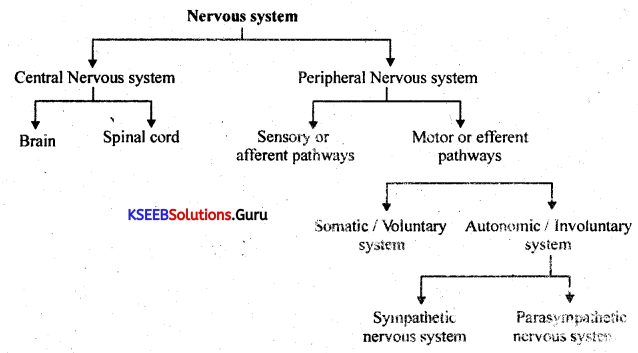
Structurally, r.iervous system is divided into 2 types. Namely,
1. Central Ne;rvous System (CNS): This includes brain and the spinal cord.
2. Peripheral Nervous System (PNS): This includes 31 pairs of spinal nerves and 12 pair of cranial herves. Cranial nerves carry impulses to and the from the brain. Spinal nerves carry impulses to and from the spinal cord. PNS is further divided into two parts, depending on the direction in which they carry impulses.
a. Afferent (Sensory) System : Consists of the sensory nerve pathways, which carry information from the receptors distributed all over the body, towards the central nervous system.
b. Efferent (Motor) system : Consists of the motor nerve pathways, which carry information away from the central nervous system to the motor organ, namely muscles and glands.
![]()
The motor system is further divided into two:-
i. Somatic Nervous System (SNS) : They carry information to the skeletal muscles. This system is said to be voluntary (This is under the control of our will)
ii. Autonomic Nervous System : They cany information to the smooth cardiac muscles and glands. This system is said to be involuntary [They work independently] and is of two types.
(a) Sympathetic Nervous System: This prepares an individual to overcome emergency situations involving fear, fight and fright.
(b) Parasympathetic Nervous System: It prepares an individual during normal conditions (non-emergency situations). It works antagonistically to sympathetic nervous system.
4. Types of Neurons:
- Neurons or nerve cells are the functional units of the nervous system.
- Groups of neurons in the central nervous system are called nuclei.
- Groups of neurons in the peripheral nervous system are called ganglia.
- Multi polar nerve cells have many short dendrites and one long axon e.g., pyramidal cells in cerebral cortex.
- A bipolar nerve cell has a long axon and one dendron, e.g., bipolar neurons in the retina.
- Unipolar nerve cells have the cell body in a side branch of the main axon; e.g., they are found in the embryonic stage.
- Present surrounding the neurons, are special companion cells known as glial cells which provide nutrition to neurons, consume their waste products and also insulate the neurons.
- Schwann cells are a type of glial cells, that wrap around the axons of neurons in the peripheral nervous system.
- A nerve has a tough covering called epineurium and on its inside, the nerve fibres are gathered into bundles (fascicles), wrapped in the perineurium.
5. Structure of Neurons:
- Each neuron has a cell body, dendrons (dendrites) and an axon.
- The cell body contains cytoplasm with a nucleus, and certain granular bodies, called Nissil granules.
- A number of processes (outgrowths) arise from the cell body. The longest among them is called axon while the others are called dendrons and their branches dendrites.
- The axon is a long fibre and is branched at its distal end. Each branch terminates as a bulb-like structure called synaptic knob, which contains synaptic vesicles with neurotransmitters.
- The axon transmits the impulses away from the cell body, while the dendrites/ dendrons conduct it to the cell body.
- Axons are of two types depending on the presence or absence of a myelin sheath around it. They are called myelinated axons/nerve fibres and non myelinated axons.
- The myelinated nerve fibres are enveloped with Schwann cells, which form a myelin sheath around the axon.
- The myelin sheath is not continuous and the gaps are called nodes of Ranvier.
- Myelinated nerve fibres are found in the brain and spiral cord, while nonmyelinated fibres are commonly found in autonomous and somatic neural systems.
![]()
6. Generation And Conduction Of Nerve Impulses:
- Neurons are excitable, as their membranes are in a polarised state.
- The neural membrane has different types of ion-channels and these ion channels are selectively permeable to different ions. This creates potential differences and is responsible for the generation and conduction of their impulses.
(i) Resting Membrane Potential:
At the resting stage i.e., when the neuron is not involved in the conduction of impulses, the membrane of neuron is more permeable to K+ ions than to Na+ ions. The membrane is impermeable to the negatively charged proteins of the axoplasm and also Cl– ions.
So the axoplasm (interior) of the axon has a high concentration of negatively charged proteins, Cl– ions and potassium ions but a low concentration of Na+ ions.
Contrary to this, the extracellular fluid (exterior) has a high concentration of Na+ ions and low concentration of K+ ions.
These ionic gradients across the membrane, are maintained by the active transport of ions by the sodium-potassium pump, which transports three Na+ outwards and two K inwards.
– So the outer surface of the axonal membrane has a positive charge and the inner surface of the membrane/axoplasm is negatively charged the membrane is said to be polarised.
(ii) Action Potential:
When a threshold stimulus is applied at a site on the polarised membrane, the membrane permeability at that site changes and becomes freely permeable to Na+ ions.
![]()
As a result, there is a rapid inflow of Na+ ions and there is a reversal in the polarity of the membrane.
The interior/axoplasm now becomes positively charged while the exterior becomes negatively charged and this is called depolarisation.
The potential difference across the membrane at the site of stimulation is known as action potential.
Now the current flows through the axoplasm from the depolarised region to the depolarised region.
As a result the action potential is generated in the next segment and this process continues along the length of the nerve fibre.
(iii) Repolarisation
At the site of stimulation, the stimulus-induced permeability to Na+ ions is extremely short lived is quickly followed by an increase in the permeability to K+ ions.
Withm a fraction of a second, K+ diffuses outside and restores the resting potential of the membrane at the tike site of excitation.
At this stags, the membrane is said to be repolarised, the next polarised region and through the ECF from Use polarised region to the depolarised region.
As a result, the action potential is generated in the next segment and this process continues along the length of the nerve fibre.
7. Transmission Of Impulses Across The Synapse:
A synapse is the functional junction between two neurons, formed by the membranes of a pre-synaptic neuron and a post-synaptic neuron.
There are two types of synapses namely:
(i) Electrical synapses and
(ii) Chemical synapses.
The differences between the two types of synapses are given below:
Electrical Synapses:
(i) The membranes of the pre-synaptic and post synaptic neurons are in close proximity
and there is no synaptic cleft.
(ii) Electrical current can flow directly from one neuron to the other.
(iii) Impulse conduction is faster.
(iv) Electrical synapses are rare in the human system.
![]()
Chemical Synapses:
(i) The membranes of the pre-synaptic and post-synaptic membranes are separated by a fluid-filled called space the synaptic cleft.
(ii) Transmission involves chemicals called neurotransmitters.
(iii) Impulse conduction is relatively slower.
(iv) Chemical synapses are the most common type of synapses in human.
In the chemical synapses, the axon terminal called synaptic knob contains a number of synaptic vesicles in its axoplasm.
The synaptic vesicles contain the neurotransmitter.
When an impulse arrives at the axon terminal, it stimulates the movement of the synaptic vesicles towards the membrane, where they fuse with the membrane and release the neurotransmitter into the synaptic cleft.
The released neurotransmitter molecules bind to their specific receptors present on the post-synaptic membrane.
This binding stimulates the opening of ion channels that can generate the action potential in the postsynaptic neuron.
8. The Human Brain:
- The brain of adult human weighs about 1.4 kg.
- The human brain is located in a protective bony cranium.
- It is externally covered by Meninges, made of a thick outer layer called duramater lying beneath the cranium, middle fibrous layer called arachnoid and the inner vascular layer called piamater.
- In between arachnoid and piamater, there is a subarachnoid space filled with cerebrospinal fluid (CSF). It protects the brain and spinal cord against mechanical injury and shocks. It also helps in exchange of nutrients, respiratory gases and metabolic wastes. It is secreted by choroid plexus.
The brain can be differentiated into three regions-fore brain, mid brain and hindbrain.
I. Fore Brain (Procencephalon) : Includes cerebrum and diencephalon (Thalamus and Hypothalamus)
II. Mid Brain (Mecencephalon) : It is a highly reduced part in humans.
III. Hindbrain (Rhombencephalon) : It includes cerebellum, ponsveroli and medulla oblongata (Stalk of the brain).
1. Fore Brain (Prosencephalon):
Cerebrum: It is the largest part of the human brain and is called greater brain.
The surface area shows numerous infoldings or convolutions. The upfolds are called Gyri (Gyrus) and deep down folds are called Fissures and shallow folds are called Sulci (Sulcus).
The cerebrum is divided into right and left cerebral hemispheres by longitudinal fissures. However, they are internally connected by a thick bundle of nerve fibres called corpus callosum. Extension of the duramater called Flax cerebri separates the two hemispheres of cerebrum.
The substance of the cerebrum can be divided into an outer cerebral cortex and an inner cerebral medulla. Cerebral cortex is packed with cell bodies and dendrites of neurons. It forms the grey matter of brain. Cerebral medulla consists of myelinated nerve fibres. It forms the white matter.
Each cerebral hemisphere is divided into four lobes, namely, frontal, parietal, temporal and occipital lobes by three deep and wide fissures.
- Central sulcus (the fissure of Rolando) separates frontal lobe from the parietal lobe.
- The lateral cerebral sulcus separates temporal lobe from the frontal lobe.
- The parieto-occipital sulcus separates parietal lobe from the occipital lobe.
![]()
Functions:
The cerebrum is the seat of intelligence, memory, consciousness, vision, speech and voluntary muscular movements.
Thalamus : It is a large oval structure present at the centre of fore brain.
Functions : It serves as a relay station that interprets sensory impulses and then channels them to the appropriate regions of the cerebral cortex.
Hypothalamus : It is present below the thalamus and largely consists of grey matter.
Functions:
a. Controls and integrates autonomic nervous system. Hence it is called head of the autonomous nervous system.
b. Regulation of body temperature.
c. Regulation of water and electrolyte balance.
d. Control of eating and drinking (hunger and thirst) through Satiety centre, and thirst centre.
e. Feeling of rage and aggression.
f. Regulation of sleep and wakefulness.
g. Regulation of heart rate and blood pressure.
h. It controls the release of honnones of anterior pituitary.
i. It secretes oxytocin and vasopressin hormones that are stored and released from posterior pituitary.
II. Mid Brain (Mecencephalon):
This comprises of optic lobes that occurs as two pairs of corpora quadrigemina.
Functions:
The corpora quadrigemina are the reflex centers of eye and auditory responses. They also control eye ball movement in response to changes with the movement of head.
III. Hindbrain:
It consists of the following three parts:-
1. Cerebellum :
- Cerebellum is the second largest portion of the brain and is called as lesser brain.
- It consists of a central constricted area called vermis and lateral wing like right and left cerebral hemispheres.
- The surface of the cerebellum consists of grey matter and is thrown into a series of parallel sulci.
- Beneath the grey matter there is a white matter and the white matter exhibits a tree like pattern called Arbor vittae.
![]()
Functions :
a. Maintenance of body posture and equilibrium.
b. Regulates certain voluntary movements that are initiated by cerebral cortex and also involved in subconscious reflexes.
2. Pons varoli :
It lies just above the medulla and acts as a bridge carrying ascending and descending tracts between brain and spinal cord.
![]()
Functions :
It is a controlling centre for salivation, mastication and facial expression etc.
3. Medulla Oblongata :
It is the posterior most part of the brain and continues into the spinal cord. The medulla contains Vital control centres for cardiac, respiratory and vasomotor centres.
Functions :
1. Contains the cardiac and respiratory centers, and controls the heart rate and respiratory rate.
2. Contains vasomotor centers that control reflexes like sneezing, coughing and hiccough etc.
3. It is a point of origin for several cranial nerves.
Note: Mid brain, ponsveroli and medulla oblongata together form brain stem.
Functions of various Topographical Areas of Cerebrum :
Cerebral cortex is broadly divided into three areas, based on the functions it controls:
1. Motor areas : Transmit impulses and control voluntary movements of various organs of the
2. Sensory areas : Receive sensory impulses and interpret them like what we see, hear, feel smell or taste.
3. Association areas : Associated with memory, learning, reasoning and intelligence.
![]()
Functions :
1. It serves as two-way conduction system as it carries stimulus from the-body tissues to the brain and from brain to various parts of the body.
2. It also carries out reflex action.
Reflex action is an automatic response to any kind of stimulus without conscious involvement of brain.
Reflex arc is the nerve pathway involved in reflex action.
It involves following components.
a. A receptor organ : It is a sensory cell where the stimulus is received.
b. A sensory neuron : It lies in the ganglion of the dorsal roof of the spinal cord, and conducts the impulses from receptor to the CNS.
c. An associated neuron : It lies in the spina! cord. It serves to transmit the impulse from the sensory neuron to the motor neuron.
d. A motor neuron : It lies in the ventral horn of the spinal cord and serves to transmit the impulse to the effector organ.
e. Effector organ : It is a muscle or gland, which responds to the impulse received.
![]()
Note : There are 12 pairs of cranial nerves and 31 pairs of spinal nerves in human.