Karnataka 1st PUC Biology Question Bank Chapter 7 Structural Organisation in Animals
1st PUC Biology Structural Organisation in Animals One Marks Questions and Answers
Question 1.
Which epithelium lines the Bowman’s capsule ?
Answer:
Squamous epithelium.
Question 2.
What type of epithelium lines your skin ?
Answer:
Squamous epithelium.
Question 3.
Which epithelium is also known as pavement epithelium ?
Answer:
Squamous epithelium.
![]()
Question 4.
Name the epithelium present on the surface of oral cavity ?
Answer:
Non-Keratinised squamous epithelium.
Question 5.
What is endothelium?
Answer:
Squamous epithelium lining the blood vessels.
Question 6.
What is mesothelium?
Answer:
Squamous epithelium lining the abdominal cavity.
Question 7.
What are goblet cells?
Answer:
Mucous secreting columnar epithelial cells.
Question 8.
What is an unicellular gland?
Answer:
A single cell secreting secretions or a goblet cell.
Question 9.
What is chondrocytes?
Answer:
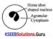
It is the adipose tissue present below the skin of whales.
Question 10.
What is chondrin?
Answer:
It is the matrix of the cartilage.
Question 11.
Where do you find haversian system ?
Answer:
In the mammalian bones.
![]()
Question 12.
What is sarcolemma?
Answer:
It is the plasma membrane covering the muscle fibre.
Question 13.
What is sarcoplasm?
Answer:
The cytoplasm present in muscle fibers.
Question 14.
What are myofibrils?
Answer:
Myofibrils are the longitudinal protein bands of muscle fibers.
Question 15.
Name the long unbranched, cylindrical, multinucleate muscle fiber?
Answer:
Skeletal muscle.
Question 16.
Where are intercalated discs present?
Answer:
Intercalated discs are present in the cardiac muscle.
Question 17.
Name the principal contractile proteins of the muscle tissue?
Answer:
Actin and Myosin.
Question 18.
Name the muscle which is striated and involuntary?
Answer:
Cardiac muscle.
Question 19.
Name the structural and functional unit of the nervous system.
Answer:
Neurons.
Question 20.
What is a cyton ?
Answer:
Cyton is the cell body of neurons having nucleus. Nissil granules, etc.
Question 21.
Where are Nissil bodies present ?
Answer:
In the cyton of neuron.
Question 22.
What is the advantage of myelin sheath ?
Answer:
a. It speeds up the conduction of nerve impulse.
b. It insulates and maintains the axon.
![]()
Question 23.
What are glial cells ?
Answer:
Glial cells are the non-nervous cells that protect and support the neurons.
or
The supporting cells of the Central Nervous System (CNS) are called Glial cells.
Question 24.
Give the binomial nomenclature of cockroach.
Answer:
The cockroach belongs to genus periplaneta and the species called americana.
Thus its binomial nomenclature is Periplaneta americana.
Question 25.
What are wing covers?
Answer:
In cockroaches there are two pairs of wings. The hindwings are thin and helpful for flight. The first pair on forewings are thick and cover the second pair while the animal is at rest. Thus they are called wing covers or tegmina.
Question 26.
What is a spiracle?
Answer:
Spiracle is an opening leading into respiratory tubes called trachea in cockroaches. The spiracle or stigmatal aperture is valvular and lateral in position.
Question 27.
What are trachea?
Answer:
Trachea are respiratory tubes in many arthropods. They branch inside to form trachioles. Trachea communicates outside through the stigmata.
Question 28.
What are alary muscles?
Answer:
The pericardial membrane in a cockroach is connected with the terga of each segment by a pair of alary muscles. These help to maintain flow of blood from haemocoel into pericardial cavity.
Question 29.
What is a phallic gland?
Answer:
A phallic gland is a club shaped gland in male reproductive system of cockroach. It is also called as conglobate body.
Question 30.
What are colleterial glands?
Answer:
In female cockroaches, there are a pair of branched whitish glands Called colleterial glands. They secrete material for egg case.
![]()
Question 31.
What is a spermatophore?
Answer:
Bundles of sperms are called spermatophores.
Question 32.
What is a ovipositor?
Answer:
Gonapophyses in females helping in deposition of eggs into the egg case are called ovipositors.
Question 33.
What is copulation?
Answer:
Copulation is an act during which sperms from the male of the species are transferred into the female body. It is also called mating.
Question 34.
What is an ootheca?
Answer:
Ootheca is the egg case of cockroach. It contains sixteen eggs arranged into two rows of eight each. This oblong, purse like egg case is formed by the secretion of colleteral glands.
Question 35.
What is a nymph?
Answer:
Nymph is the stage in the development of cockroach. It resembles an adult except for wings and gonads.
Question 36.
What is incomplete metamorphosis?
Answer:
Changes in structure and function leading to the transformation of immature form of young into an adult is called metamorphosis. Nymph resembels the adult but not fully, and therefore the changes are minor. This type of metamorphosis is called incomplete matamorphosis.
Question 37.
What are palpigers?
Answer:
Spongy cushion like structures at the base of labiale palps in cockroaches/or arthopodes are called palpigers.
Question 38.
What are fenestrae?
Answer:
Fenestrae are reduced, functionless eyes or ocelli of cockroaches.
Question 39.
Which is the strongest segment in the leg of cockroach?
Answer:
Femur (the third) is the strongest segment in the cockroach leg.
Question 40.
What are the structures found between midgut and hindgut?
Answer:
Malpighian tubules occur between midgut and hindgut. They are thin, hairlike, yellowish excretory bodies.
![]()
Question 41.
Answer in one word or one line.
(i) Give the common name of Periplaneta americana.
Answer:
American Cockroach.
(ii) How many spermathecae are found in an earthworm?
Answer:
Two.
(iii) What is the position of ovaries in cockroach?
Answer:
Dorsolaterally in 4-5 abdominal segments.
(iv) How many segments are present in the abdomen of a cockroach?
Answer:
Ten.
(v) Where do you find Malphigian tubules?
Answer:
At the junction between midgut and ileum part of hindgut in insects.
Question 42.
Mention the functions of the following
(a) Ureters in a frog.
(b) Malpighian tubules.
(c) Body wall in earthworm.
Answer:
(a) Act as urinogenital duets
(b) Excretion
(c) Protection.
Question 43.
Where are the sclerites present in a cockroach?
Answer:
In the exoskeleton.
Question 44.
Which mouth part of cockroach is comparable to our tongue?
Answer:
Hypopharynx(= lingua)
Question 45.
Give the location of hepatic caeca in a cockroach. What is their function?
Answer:
Location : Hepatic caeca are a ring of 6-8 blind tubules found at the junction of foregut and midgut. Their function is to produce the digestive enzymes.
Question 46.
In which segment of the earthworm, female genital pores are present?
Answer:
14th segment.
Question 47.
In which segment of the earthworm, male genital pores are present?
Answer:
18th segment.
Question 48.
What is Vasa deferentia?
Answer:
It is the common prostrate and spermatic duct.
Question 49.
On which segments of the earthworm two pairs of testes occur?
Answer:
10 the and 11th segments.
Question 50.
On which segments of the earthworm a pair of ovaries occur?
Answer:
12th and 13th segments.
![]()
Question 51.
What is the food of an earthworm?
Answer:
It is the decaying leaves and organic matter mixed with soil.
Question 52.
What are Nephridia?
Answer:
They are the excretory organs in earthworm.
Question 53.
What are sclerites?
Answer:
Each segment of the exoskeleton, of a cockroach has hardened plate like subunits called as sclerites.
Question 54.
What is Haemocoel?
Answer:
It is the blood filled body cavity in a cockroach.
Question 55.
How many chambers are there in heart of cockroach?
Answer:
13 chambers.
Question 56.
How many pairs of spiracles are present in the heart of cockroach?
Answer:
10 pairs.
Question 57.
What is Phallomere?
Answer:
It is the chitinous asymmetrical structure surrounding the male genopore in a cockroach.
Question 58.
What are spermatophores?
Answer:
In cockroaches, sperms stored in the seminal vesicles are glued together in the form of bundles called spermatophores.
Question 59.
How many ovarioles are present in the ovary of a cockroach?
Answer:
Eight ovarioles.
Question 60.
How many ganglia occur in the thorax of a cockroach?
Answer:
Three ganglia.
Question 61.
What is Nymphs?
Answer:
Young ones of a cockroach are called Nymphs.
Question 62.
What is Aestivation?
Answer:
It is the summer sleep in frog.
Question 63.
What is Hibernation?
Answer:
It is the winter sleep in frog.
Question 64.
Name the membrane in frogs, which receives sound signals?
Answer:
Tympanum.
1st PUC Biology Structural Organisation in Animals Two Marks Questions and Answers
Question 1.
Write any four main characteristic features of epithelial tissues.
Answer:
The four main characteristic features are:-
a. The cells are arranged compactly, without intercellular spaces.
b. Epithelial cells always rest on the basement membrane composed of fine fibers and non living protein polysaccharide material.
c. The cells are arranged in the form of a continuous layer or sheet. Such a sheet may cover the external surface of an organ or internal cavity.
d. The cells may be grouped as tubes or follicles to produce glands.
e. Epithelial tissues are avascular.
![]()
Question 2.
State any four functions of epithelium.
Answer:
The four functions of epithelium are,
a. Protection: The epithelial tissues protect the underlying tissue from being dried out.
b. Absorption: The lining of the intestine absorbs food, and the lining of the kidney tubules selectively absorbs certain substances.
c. Transport: The materials absorbed by the epithelium is transported across them either to blood or to lymph.
d. Secretion: Epithelial cells either in unicellular or multicellular states, secrete substances.
Question 3.
Bring out any four differences between cartilage and bone.
Answer:
| Cartilage | Bone |
| 1. It is restricted to few organs like ear pinna, nose tip, larynx and is the minor component of endoskeleton. | 1. It occurs throughout the body of the vertebrates and is the major component of endoskeleton. |
| 2. Cartilage is covered by a membrane called perichondrium. | 2. Bone is covered by a membrane called periosteum. |
| 3. Lacunae are distributed randomly. | 3. Lacunae are arranged in concentric layers. |
| 4. Cells are known as chondrocytes. | 4. Cells are known as osteocytes. |
| 5. The matrix is chondrin | 5. The matrix is ossein. |
Question 4.
Write any four distinctive features of a connective tissue.
Answer:
a. The cells are not grouped but are spaced at some distance from one another i.e., intercellular spaces are present.
b. The intercellular spaces are filled up by the material produced by some cells which forms ground substance.
c. The tissues are vascularised and innervated.
d. They are derived from mesoderm.
Question 5.
State any four differences between epithelial and connective tissues.
Answer:
| Epithelial tissue | Connective tissue |
| 1. The cells are arranged compactly i.e., intercelluar spaces are absent. | 1. The cells are not grouped but are spaced at some distance from one another i.e. intercelluar spaces are present. |
| 2. The epithelial cells rest on basement membrane. | 2. The connective tissue cells do not rest on basement membrane. |
| 3. The cells are arranged in the form of a continuous laver or sheet. | 3. The cells are not arranged in any form of continuous layer or sheet. |
| 4. Epithelial cells are avascular. | 4. The cells are vascular. |
Question 6.
Mention any four functions of a connective tissue.
Answer:
a. They help in cementing between the tissues.
b. They help as packaging tissues.
c. They help in the deposition of the fat on the surface of the body.
d. They provide protection and cushioning effect to the vital internal organs.
Question 7.
Write a note on the matrix of bone.
Answer:
The ground substance of bone tissue is amorphous and consists of both organic and inorganic components. The organic components are glycoproteins and mucopolysaccharides which form osteoid. The matrix consists of calcium phosphate and calcium carbonate and hence it is hard. The matrix occurs in the form of rings. Each ring or layer is called a lamella. They are arranged concentrically to’form a central tubular passage called haver sian canal.
Question 8.
Write a note on white fibers.
Answer:
White fibers are also called collagen fibers. They are composed of bundles of smaller fibers of protein called collagen. They are flexible but tough and serve to give strength to the tissue. They do not show branching.
Question 9.
Mention two structural dissimilarities between cardiac and striated muscle.
Answer:
| Cardiac muscle | Striated muscle |
| 1. They are uninucleated. | 1. They are multinucleated. |
| 2. Muscle fibers are branched and these run parallel!. Intercalated discs are present. |
2. Muscle fibers are unbranched. Intercalated discs are absent. |
Question 11.
Mention two similarities between striated and cardiac muscle.
Answer:
| Striated Muscle | Cardiac muscle |
| 1. They are cylindrical with blunt ends. | 1. They are also cylindrical in structure. |
| 2. Cross bands are present. | 2. Cross bands are present. |
![]()
Question 12.
Mention a few dissimilarities between smooth and cardiac muscles.
Answer:
| Smooth muscle | Striated muscle |
| 1. They are spindle-shaped with pointed ends; | 1. They are cylindrical with blunt ends. |
| 2. Cross bands are absent however longitudinal myofibrils are present. | 2. Cytoplasm shows the alternate bright and dark bands. |
| 3. They are uninucleated. | 3. They are multinucleated. |
| 4. They are involuntary. | 4. They are voluntary. |
| 5. They do not suffer fatigue. | 5. They suffer fatigue. |
Question 13.
Draw a labelled sketch of a smooth muscle cell.
Answer:
Question 14.
Give four differences between smooth and striated muscles.
Answer:
| Smooth muscle | Striated muscle |
| 1. It occurs in visceral organs. | 1. It occurs in neck, limbs trunk, etc. |
| 2. Spindle shaped with pointed ends. | 2. Cylindrical with blunt ends. |
| 3. They are uninucleated. | 3. They are multinucleated. |
| 4. Longitudinal myofibrils | 4. Alternate cross bands are present. |
Question 15.
Name the different sensory organs in the head of a cockroach?
Answer:
In the head of a cockroach, there are sense organs like:
(1) Compound eyes.
(2) Ocelli (degenerated eyes).
(3) Antennae.
(4) Sensory bristles on the mouth parts.
Question 16.
Give the classification of cockroach.
Answer:
Kingdom : Animalia.
Phylum : Arthropoda.
Class : Insecta.
Order : Dictyoptera.
Family : Blattidae.
Genus : Periplaneta.
Species : americana:
![]()
Question 17.
Answer the following:
(i) What is the function of nephridia?
Answer:
It is the excretory organ of the members belonging to annelida.
(ii) How many types of nephridia are found in an earthworm, based on their locations?
Answer:
There are 3 types of nephridia:
(a) Septal nephridia – Being is the segment.
(b) Integumentary nephridia – Inner side of body wall in all segment expects.
(c) Pharyngeal nephridia – 4th, 5th and 6th segments.
Question 18.
What are the cellular components of blood?
Answer:
RBC, WBC and platelets.
Question 19.
Comment on the structure of Clitellum.
Answer:
In a mature earthworm, segments 14th, 15th and 16th are covered by a prominent dark band of glandular tissue called clitellum. Based on this structure, body of an earth worm is divisible into three regions such as pre-clitellar, clitellar and post clitellar segments Clitellum does not bear setae.
Question 20.
What is typhlosole? Give its function.
Answer:
In an earthworm, as a characteristic feature of the intestine, between 26th to 35th segments there is the presence of an internal median fold of dorsal wall called typhlosole. It helps in increasing effective area for absorption in the intestine.
![]()
Question 21.
Where are the blood glands present in an earthworm? Give their function.
Answer:
Blood glands are present on the 4th, 5th and 6th segments. They produce blood cells and haemoglobin which is dissolved in plasma.
Question 22.
Why earthworms are called “friend of formers”?
Answer:
Earthworms are called friend of farmers because they make burrows in the soil and make it porous which helps in respiration and penetration of developing plant roots. Fertility of soil is also increased by their vermicompost.
Question 23.
What is mosaic vision? Describe.
Answer:
With the help of several ommatidia, a cockroach can receive several images of an object. This kind of vision is known as mosaic vision. With more sensitivity but less resolution, being common during night, it is also called nocturnal vision.
1st PUC Structural Organisation in Animals Three Marks Questions and Answers
Question 1.
Complete the following statements:
(a) In a cockroach, grinding of food particles is performed by
(b) Malpighian tubules help in. the removal of
(c) Hindgut of cockroach is differentiated into
Answer:
(a) gizzard / proventriculus.
(b) nitrogenous waste products.
(c) ileum, colon and rectum.
Question 2.
Write the appropriate type of tissues in column B according to the functions mentioned in column A.
Column A —- Column B
(a) Secretion and absorption — ……………………
(b) Protective covering — ……………………
(c) Linking and supporting framework — ……………………
Answer:
(a) Columnar epithelium.
(b) Stratified epithelium.
(c) Connective tissue.
![]()
Question 3.
Classify and describe the epithelial tissue types on the basis of structural modification of cells.
Answer:
1st PUC Biology Structural Organisation in Animals Five Marks Questions and Answers
Question 1.
What is an epithelial tissue ? Describe any two types of simple epithelial tissues.
Answer:
Epithelial tissue is a tissue, which lies on the upper and outer layer covering surface of the organ or lining the cavity.
Squamous epithelium:
a. It occurs in the skin of vertebrates. Alveoli of lungs, the mouth cavity, etc.
b. The cells are nearly hexagonal flat and plate like.
c. A spherical or oval nucleus is found to be present at the cell centre.
d. The cells are closely fitted with minimum intercellular space. Hence they look like a mosaic pavement. Because of this, squamous epithelium is also called as pavement epithelium.
e. It forms a continuous cover, and protects the inner tissues.
Columnar epithelium:
a. It occurs in the mucosa membrane of the stomach and the intestine.
b. The cells are elongated and pillar like.
The lower ends are narrow and upper ends are broad.
c. The nucleus is oval and basal cytoplasm is granular.
d. Cells are lodged over a basement membrane. Intercellular spaces are present at the basal region of the cells.
e. The columnar cells are absorptive and protective in function.
Question 2.
With a neat labelled sketch, explain the structure of areolar tissue.
Answer:
a. It is present beneath the skin. It is also known as fatty tissue.
b. Adipose tissue is made up of two components, namely.
(i) Matrix: It is also called ground substance. It is a homogenous substance. It contains cells and blood vessels.
(ii) Cells: Cells of the adipose tissue are called adipocytes or fat cells as they contain large amount of fat enclosed in a pcotoplasmic sheath. They are spherical or oval in shape ‘Vacuolc
and lie close to blood vessels. The nucleus Nuckus and cytoplasm are pushed towards the periphery.
c. Sometimes between these cells, fibres may be present but they are ‘sparse’.
d. In whales, adipose tissue forms a layer all around the body calledithibber it is insulatory and storage in function.
![]()
Question 3.
Draw a labelled diagram of hyaline cartilage and describe the parts.
Answer:
Question 4.
Write a neat labelled diagram and explain the structure. of the haversian system.
Answer:
Question 5.
Draw a labelled diagram of the reproductive organs of an earthworm.
Answer:
Question 6.
Draw a labelled diagram of the alimentary canal of a cockroach.
Answer:
Female Cockroach:
I. Abdomen broad and short.
2. Anal styles absent.
3. A brood pouch enclosed in the 7th, 8th and 9th sterna ¡s present
4. Antennae shorter.
5. Seventh abdominal sterna are visible only.
6. Only the first seven abdominal sterna are visible (only).
7. Genital aperture lies on the 8th sternum.
8. Gonapophyses consists of pairs of plates,
Male Cockroach:
1. Abdomen narrow and long.
2. Anal styles present. They are in the form of unjointed appendages attached to the 9the segment.
3. No brood pouch.
4. Antennae longer.
5. Seventh abdominal tergum covers only the 8th sternum.
6. All the nine abdominal sterna are visible.
7. Gential aperture lies below the anus between the 10th tergum and 9th sternum.
8. Gonapophyes consists of 3 phallomeres only.

![]()
Question 7.
Describe various types of epithelial tissues, with the help of labelled diagrams.
Answer:
Classification of epithelial tissues:
Epithelial tissues are broadly classified into two types namely:
(a) Simple epithelium: Single layered epithelial tissue is known as simple epithelium.
(b) Stratified epithelium: Multilayered epithelial tissue is known as stratified epithelium.
(a) Simple epithelium: Simple epithelium is further classified into:
(i) Squamous epithelium.
(ii) Columnar epithelium.
(iii) Cuboidal epithelium.
(iv) Ciliated epithelium.
(v) Glandular epitheliuni
(i) Squamous epithelium:
1. It occurs in the skin of vertebrates, alveoli of lungs, the mouth cavity. etc.
2. The cells are nearly iexagonal, flat and plate like.
3. A spherical or oval nucleus ¡s found at the cell centre.
4. The cells are closely fitted without intercellular space. Hence look like a mosaic pavement.
Because of this squamous epithelium is also called pavement epithelium.
Function:
1. It forms a continuous cover and protects the inner tissues.
2. It helps in ultrafiltration in Bowman’s capsule.
3. It helps in exchange of gases in alveoli.
(ii) Columnar epithelium:
1. It occurs in the mucosa layer of stomach and intestine.
2. The cells are elongated and pillar like. The lower ends are narrow and upper ends are broad.
3. The nucleus is oval and basal and cytoplasm is granular.
4. Cells are lodged over a basement membrane. Intercellular spaces are present at the basal region of the cells.
5. The free surface of the cells may be smooth or branched to form microvilli.
Function:
The columnar cells are absorptive and protective in function
(iii) Cuboidal epithelium:
1. Cuboidal epithelium occurs in organs like thyroid, kidney, etc.
2. The cells are cuboidal in shape with central spherical nucleus.
(iv) Ciliated epithelium:
1. It occurs in the trachea of the lungs and the urinogenital ducts (oviduct, ureter).
2. The cells are elongated and pillar like.
3. The lower end of the cell is attached to basement membrane and the upper end is free. The upper border bears hair like cilia.
Question 8.
Distinguish between,
(a) Simple epithelium and compound epithelium.
(b) Cardiac muscle and striated muscle.
(c) Dense regular and dense irregular connective tissues .
(d) Adipose and blood tissue.
(e) Simple gland and compound gland.
Answer:
Smooth muscle or non-striped muscle:
1. It occurs in visceral organs like stomach, intestine. etc.
2. The fibre occurs in sheaths or bundles. Each fibre is narrow and spindle shaped.
3. Nucleus is oval and lies at the centre
4. The cytoplasm is called sarcoplasm and does not show cross bands. The sarcoplasm contains longitudinal myofibrils that cause contraction.
5. The contraction of smooth fibres is slow and not controlled by will, hence they are called involuntary muscles.
6. They do not suffer fatigue.
![]()
Cardiac muscle:
1. It is found only in the heart of vertebrates. Hence called cardiac muscle.
2. The cardiac fibres are arranged lengthwise with little connective tissue in between.
3. The fibres are joined at the ends by modified sarcolemma called intercalated disc.
4. Each fibre has a oval nucleus at the centre. The fibres show less distinct cross bands.
5. The fibres show free lateral branches or lateral processes, which establish connection with adjacent fibres.
6. The function of cardiac muscles is not under the control of will, hence called involuntary muscle. They do not suffer from fatigue.
Question 9.
Mark the odd one, in each of these series:
(a) Areolar tissue; blood; neuron; tendon
(b) RBC; WBC; platelets; cartilage – Blood /bone.
(c) Exocrine; endocrine; salivary gland; ligament – glandular /Tissue.
(d) Maxilla; mandible; labrum; antennae – mouth parts/ feeler.
(e) Protonema; mesothorax; metathorax; coxa
Answer:
(a) Neuron
(b) Cartilage
(c) Ligament
(d) antennae
(e) Protonema.
Question 10.
Match the terms in column I with those in column II:
Answer:
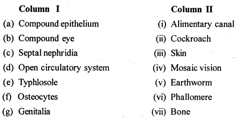
Answer:
(a) – (iii), (b) – (iv), (c) – (i), (d) – (ii), (e) – (v). (f) – (vii), (g) – (vi).
Question 11.
Mention briefly about the circulatory system of earthworm.
Answer:
Excretory System: Excretion takes place by means of a large number of coiled tubular structures known as nephridia. These are the excretory units. There are 3 types of nephridia found in earthworm.
(a) Integumentary Nephindia: These are exonephric, present on the inner side of the body wall in all the segments except the first two segments. Their number is 200 to 250 in each segment except in clitellum, where they are 10 times more. These are known as “Forests of nephridia.
(b) Septal Nephndia : These are enteronephric and sitijated behiñd the 15th segment attached to the septa, on both sides. Each row has 80 to 100 septal nephredia. They are enteronephric.
![]()
(c) Pharyngeal Nephridia: They are enteronephric. Three groups of pharyngeal nephridia are situated in each of the 4th, 5th and 6th segments. Out of all the 3 types of nephridia,
only septal nephridia have a funnel-shaped structure known as nephrostome and thus can collect tbe waste products from the coelomic fluid, other two types don’t have nephrostome. .
Question 12.
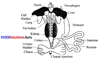
Draw a neat diagram of the digestive system of a frog.
Answer:
1st PUC Biology Structural Organisation in Animals Text Book Questions and Answers
Animal Histology:
Group of cells having similar origin, structure and function is called a tissue. Tissue is a group of closely associated cells adapted to carry out a specific function or a set of functions. Branch of biology which deals with the study of tissues is called Histology. The origin of these tissues take place from the three primary germ layers namely, ectoderm, mesoderm and endoderm. The process of formation of tissues from the three germ layers of the embryo is called histogenesis.
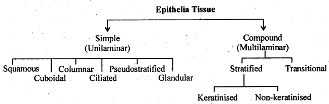
Classification of animal tissues:
Basically animal tissues are of four types, namely:
1. Epithelial tissues.
2. Muscular tissues.
3. Connective tissues.
4. Nervous tissues.
1. Epithelial Tissue:
Tissue which lie on the outer or upper layer covering the surface of an organ or lining the cavity are called epithelial tissues.
Characteristic features of epithelial tissue:
1. They form a lining both to external and internal surfaces of the body.
2. Cells are compactly arranged. As a result of such an arrangement, the intercellular substance is highly reduced.
A thin basement membrane is present usually below the epithelium.
The cells are attached to each other and to the basement membrane by means of cell membrane proteins and specialized areas of cell membrane called cell junctions.
The tissue is avascular i.e.blood vessels are absent.
Epithelial tissues are formed by all the three germ layers.
Classification of epithelial tissues:
Epithelial tissues are broadly classified into two types namely:
(a) Simple epithelium: Single layered epithelial tissue is known as simple epithelium.
(b) Stratified epithelium: Multilayered epithelial tissue is known as stratified epithelium.
(a) Simple epithelium: Simple epithelium is further classified into:-
- Squamous epithelium.
- Columnar epithelium.
- Cuboidal epithelium.
- Ciliated epithelium.
- Glandular epithelium.
Squamous epithelium :
1. It occurs in the skin of vertebrates, alveoli of lungs, the mouth cavity, etc.
2. The cells are nearly hexagonal, flat and plate like.
3. A spherical or oval nucleus is found at the cell centre.
4. The cells are closely fitted without intercellular space. Hence look like a mosaic pavement. Because of this squamous epithelium is also called pavement epithelium.
Function:
1. It forms a continuous cover and protects the inner tissues.
2. It helps in ultrafiltration in Bowman’s capsule.
3. It helps in exchange of gases in alveoli.
Note: Squamous epithelium lining the blood vessels is called endothelium( derived from endoderm). Squamous epithelium lining internal organs suah as stomach, intestine is called mesothelium (derived from mesoderm).
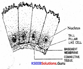
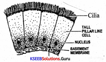
(ii) Columnar epithelium:
1. It occurs in the mucosa layer of stomach and intestine.
2. The cells are elongated and pillar like. The lower ends are narrow and upper ends are broad.
3. The nucleus is oval and basal and cytoplasm is granular.
4. Cells are lodged over a basement membrane. Intercellular spaces are present at the basal region of the cells.
5. The free surface of the cells may be smooth or branched to form microvilli.
Function:
The columnar cells are absorptive and protective in function.
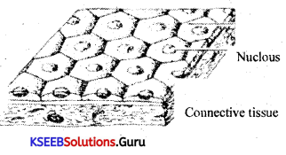
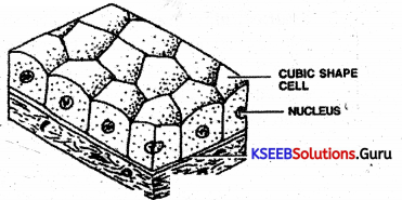
(iii) Cuboidal epithelium:
1. Cuboidal epithelium occurs in organs like thyroid, kidney, etc.
2. The cells are cuboidal in shape with central spherical nucleus.
3. These cells are lodged over basement membrane without intercellular spaces.
Function:
Cuboidal epithelium is protective in function.
![]()
(iv) Ciliated epithelium:
1. It occurs in the trachea of the lungs and the urinogenital ducts (oviduct, ureter).
2. The cells are elongated and pillar like.
3. The lower end of the cell is attached to basement membrane and the upper end is free. The upper border bears hair like cilia.
Function:
1. Cilia are protoplasmic processes which into specific direction. Hence helps to sweep fluids
2. Helps in the movement of gametes in the genital ducts.
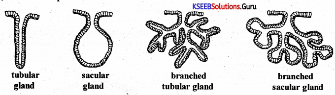
(v) Glandular epithelium:
1. They are found as isolated cells among the columnar cells as collections of the glands.
2. The cells are columnar or cuboidal and specialized for secretion.
3. Mucous secreting cells of the glandular epithelium are called goblet cells.
4. A single folding forms a sacular or a tubular gland and repeated folding forms branched gland.
Types of glands:
1. Unicellular glands: They are single secretory cells occuring amongst other non -secretory cells. They are found in the surface epithelium.
e.g: Goblet cells
Goblet cells are mucous secreting cells occuring in the wall of stomach, intestine or respiratory tract.
2. Multicellular glands: They are true glands wherein a large number of glandular cells are arranged to form a definite secretory body or gland. They are shaped up in various ways to have extra space for secretion.
e.g: Intestinal glands, pancreas etc.
Multicellular glands can be classified into simple and compound glands:
(i) Simple glands: They contain a single secretory unit. The secretory portion may be tube like when ft is tubular or it may be sac like when it is sacular or acinar or alveolar. Further the single secretory unit may be branched or coiled (in case of tubular). e.g: Sweat glands of the skin are simple tubular and coiled.
Cutaneous glands of frog are simple acinar glands
(ii) Compound gland: Those containing several secretory units combined into a gland. These can be further divided as follows.
(b) The compound sacular ( or alveolar) gland: A group of simple alveolar type associated with specific function represent compound alveolar type of gland. e.g: Alveoli of lungs, Salivary gLands.
(c) The mixed or branched gland: A gland having many secretory units which are both tubular and alveolar in appearance is a mixed gland. These are also called tub ulo – acinar glands.
e.g: Sub – maxillary gland, Sub – lingual gland, Pancreas.
Function:
Cells of glandular epithelium utilise raw materials from blood and synthesise various substances like mucous, enzymes, hormones, etc.
Pseudostratified epithelium: It is also a single layered epithelium. All the cells rest on basement membrane. However, the cell size varies. Only the tallest reach the surface. The nucleii of the cells are located at different levels giving pseudo impression of multiple layers, hence the name pseudostratified epithelium. It lines most of the respiratory passages.
(b) Stratified epithelium: As the name indicates, it is composed of strata (sing: stratum: L. Layer) of two or more cells in thickness. There are several types of stratified epithelia.
They are as shown below:-
(i) Stratified squamous epithelium: It occurs in the epidermis of the skm. In the skin the superficial cells become progressively hard due to the deposition of a protein called keratin. The process in called keratinization or comification, which kills the cell. Hence the outer most layer of cells are dead and form the stratum comium or, homy layer. Maximum comification is in the sole of foot.
The mouth cavity, tongue, pharynx, oesophagus and anal canal of humans are also lined by stratified squamous epithelium. But this is nori-keratinized or non-comified. In the skin it is keratinized.
(ii) Stratified cuboidal epithelium: It forms the sweat glands and ducts of larger glands like the salivary gland and the pancreas.
(iii) Stratified columnar epithelium: It forms the wall of the ducts of mammary gland and the urinaiy passage (urethra ) in the male.
(iv) Stratified columnar ciliated epithelium: It lines the larynx.
2. Muscular Tissue Or Contractile Tissues:
It is composed of highly modified cells with the property of contractions and elongation. Thus they bring about movements of the body. Muscle cells are also called muscle fibres. Contraction and elongation of muscle fibres is due to the presence of actin and myosin, which are called contractile proteins.
Muscle fibres are of three types. They are:
(a) Skeletal muscle
(b) Smooth muscle
(c) Cardiac muscle.
(i) Stratified squamous epithelium: It occurs in the epidermis of the skm. In the skin the superficial cells become progressively hard due to the deposition of a protein called keratin. The process in called keratinization or comification, which kills the cell. Hence the outer most layer of cells are dead and form the stratum comium or, homy layer. Maximum comification is in the sole of foot.
The , mouth cavity, tongue, pharynx, oesophagus and anal canal of humans are also lined by stratified squamous epithelium. But this is nori-keratinized or non-comified. In the skin it is keratinized.
(ii) Stratified cuboidal epithelium: It forms the sweat glands and ducts of larger glands like the salivary gland and the pancreas.
![]()
(iii) Stratified columnar epithelium: It forms the wall of the ducts of mammary gland and the urinaiy passage (urethra ) in the male.
(iv) Stratified columnar ciliated epithelium: It lines the larynx.
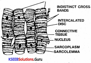
(a) Skeletal muscle or Striped muscle or Striated muscle
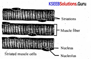
1. It is or present in limbs, neck, abdomen etc.
2. The fibres are elongated and cylindrical with blunt ends. They are parallel to one another and form bundles.
3. Each fibre is covered by a tough membrane called sarcolemma.
4. Each fibre contains many nuclei (multinucleated) and they are peripherally scattered below the sarcolemma.
5. The cytoplasm is called sarcoplasm and it shows a characteristic dark and light bands in regular alternation.
6. The fibres show rapid contractions and controlled by will, hence are cadled voluntary muscles. They suffer fatigue.
Note: utra structure of skeletal muscle fibres: The skeletal muscle cell (Fibre) is covered by thick and tough cell membrane called sarcolemma. Each fibre is multinucleate, syncytial or coenocytic. Many nuclei are scattered at the periphery of the fibre directly under the sarcolemma. The cytoplasm (sarcoplasm) contains the myofibrils, composed of myosin (forming thick filaments) and actin and other proteins (forming thin filaments) and show characteristic transverse markings. The markings together produce a regular alternation of light and dark stripes or striations or band across a fibre. Hence it is also called striated or striped fibre.
The repeating dark and light bands are seen along the length of each myofibril.
The dark bands are called A – bands (A for anisotropic) and the light are called I – bands (I for isotropic). Electron microsocpe shows that these are due to a regular arrangement of thick (myosin) and thin (actin) filaments. The centre of the A – band is lighter. It is called H – Zone ( H for hella meaning bright). The H – Zone itself is bisected by a M line.
Passing through the middle of each I – band is a dark line called Z – line (also called Krause’s membrane). The portion of a myofibril between two Z – lines is called a sarcomere. It is 2.0 micrometre long. It is the smallest contractile unit of a muscle fibre.
An individual skeletal muscle fibre and bundles of fibres are surrounded by a connective tissue. At the end of the muscle, the connective tissue is continued as the tendon.
![]()
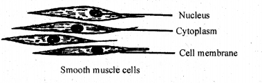
(b) Smooth muscle or non-striped muscle:
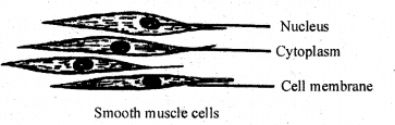
1. It occurs in visceral organs like stomach, intestine, etc.
2. The fibre occurs in sheaths or bundles.
Each fibre is narrow and spindle shaped.
3. Nucleus is oval and lies at the centre.
4. The cytoplasm is called sarcoplasm and does not show cross bands. The sarcoplasm contains longitudinal myofibrils that cause contraction.
5. The contraction of smooth fibres is slow and not controlled by will, hence they are called involuntary muscles.
6. They do not suffer fatigue.
(c) Cardiac muscle:
1. It is found only in the heart of vertebrates. Hence called cardiac muscle.
2. The cardiac fibres are arranged lengthwise with little connective tissue in between.
3. The fibres are joined at the ends by modified sarcolemma called intercalated disc.
4. Each fibre has a oval nucleus at the centre. The fibres show less distinct cross bands.
5. The fibres show free lateral branches or lateral processes, which establish connection with adjacent fibres.
6. The function of cardiac muscles is not under the control of will, hence called involuntary muscle. They do not suffer from fatigue.
Differences between striped skeletal and smooth muscles:
Skeletal muscle:
1. It occurs in neck, limbs, trunk etc.
2. Cylindrical cells with blunt ends.
3. The cells are multinucleated and nuclei are peripheral.
4. Cytoplasm shows alternate dark and light cross bands.
5. Voluntary in function.
6. They suffer fatigue.
Smooth muscle
1. It occurs in visceral organs like intestine, urinary bladder, stomach etc,.
2. Spindle shaped cells with pointed ends.
3. The cells are uninucleated and nucleus is present at the centre.
4. No such cross bands are present, however longitudinal myoffbrils are present.
5. Involuntary in function.
6. They do not suffer fatigue.
Differences between Skeletal muscle and Cardiac muscle:
Skeletal muscle:
1. It occurs in limbs, neck, trunk etc.
2. Each fibre contains a number of nuclei just below sarcolemma.
3. Muscle fibres are unbranched and run parallel.
4. Cross bands are distinct.
5. It is voluntary in function.
6. They suffer fatigue.
Cardiac muscle:
1. It occurs only in heart of vertebrates.
2. Each fibre contains nucleus at the centre.
3. Muscle fibres are branched and these branches connect the adjacent cells.
4. Cross bands are less distinct.
5. It is involuntary in function.
6. They do not suffer fatigue.
Differences between Smooth muscle and Cardiac muscle:
Smooth muscle:
1. It occurs in visceral organs.
2. The cells are spindle shaped and unbranched.
3. Cross bands are absent.
4. Intercalated discs are absent.
5. Contractions are slow and rhythmic.
Cardiac muscle:
1. It occurs only in heart.
2. The cells are cylindrical and branched.
3. Feeble cross bands are present.
4. Intercalated discs are present.
5. Contractions are rapid and rhythmic.
![]()
3. Connective Tissues:
Connective tissue includes those which bind other tissues and also supporting and packing tissues. Connective tissue is of three are types namely, connective tissue proper, Skeletal tissue and Vascular tissue.
Connective tissue proper:
Mainly it is a binding tissue. It is of two types, namely,
(A) Loose connective tissue
( B) Dense connective tissue
(A) Loose connective tissue: It has loosely distributed fibres in the organic matrix hence the name. Examples for loose connective tissue are
(1) Areolar tissue
(2) Adipose tissue.
(1) Areolar tissue: .
a. It is present beneath the skin and in between the muscle fibres.
b. Areolar tissue is made up of three major components.
(i) Matrix.
(ii) Cells
(iii) Fibres.
(i) Matrix: It is the homogenous and transparent substance. It is also known as ground substance. It contains five types of cells and two types of fibres.
(ii) Cell types :
(a) Fibroblasts or Fibrocytes: These are large and elongated cells with branched ends. They have a oval nucleus and granular cytoplasm. They secrete white and yellow fibres.
(b) Macrophages: These are large cells that move about. They swallow (ingest) foreign bodies like bacteria, thus provide on serve a defence mechanism.
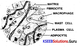
(c) Mast cells: These are spherical or oval shaped cells with spherical nucleus and cytoplasm. They secrete anticoagulatory substances like heparin. They also secrete histamine and serotonin.
(d) Adipose cells or Adipocytes: They are spherical cells and contain large quantity of lipids.
(e) Plasma cells: They are oval shaped cells with agranular cytoplasm and small nucleus.
They produce antibodies.
(iii) Fibres:
(a) White fibres: They are also called collagen fibres, as they are made up of collagen. They are tough and provide strength.
(b) Yellow fibres : They are also called elastin fibres as they are made up of elastin. They are branched and provide elasticity.
(2) Adipose tisssue :
a. It is present beneath the skin. It is also known as fatty tissue.
b. Adipose tissue is made up of two components, namely.
(i) Matrix: It is also called ground substance. It is a homogenous substance. It contains cells and blood vessels.
(ii) Cells: Cells of the adipose tissue are called adipocytes or fat cells as they contain large amount of fat enclosed in a protoplasmic sheath. They are spherical or oval in shape and lie close to blood vessels. The nucleus and cytoplasm are pushed towards the periphery.
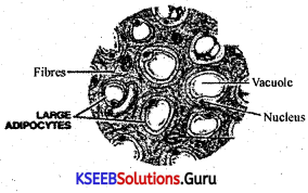
c. Sometimes between these cells, fibres may be present but they are ‘sparse’.
d. In whales, adipose tissue forms a layer all around the body called blubber. It is insulatory and storage in function.
(B) Dense connective tissue: It has closely packed fibres, hence the name, Dense connective tissue occurs in the form of sheets or chord like structures. Examples for dense connective tissue are
(i) Tendon
(ii) Ligament.
(i) Tendons: They are exclusively composed of white collagenous fibres arranged in regular bundles. In between these fibres a few fibroblasts may occur. Usually they connect muscle to bone.
(ii) Ligaments: They may contain both white and yellow fibres and these fibres are arranged in less orderly pattern. Usually they connect bone to bone.
Skeletal tissue:
Mainly it is a supportive tissue and is of two types, namely.
(a) Bone: Bone is also known as osseous tissue and it is the most rigid form of connective tissue.
Bone is not completely a solid homogenous structure. The regions of a long bone may be recognized into spongy bone (found at the ends – Epiphysis) and compact bone (found in the middle – Diaphysis).
Spongy bone may contain many large spaces filled with red marrow which is a soft tissue that produces RBC. Numerous branching structures called trabeculae are also present.
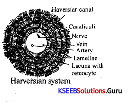
Compact bone is a solid portion of the bone. The hollow core of compact bone is called medullary cavity or marrow cavity in which yellow marrow is present. This cavity is continuous with the spaces of spongy bone. The marrow cavity is lined by endosteum. The outer surface of the bone is completely enclosed by fibrous and vascular membrane called periosteum.
In between periosteum and endosteum, matrix of the bone is present which is referred to as ossein. Ossein of mammalian bone contains number of Haversian canals or osteonic canals. These Haversian canals run parallel to the long axis of the bone and are interconnected by transverse communication canals called Volkmann’s canals. A Haversian canal with its surrounding lamellae, lacunae, osteocytes and canaliculi is called a Haversian system or Osteon.
Structure of a Haversian system:
1. Bone is composed of a number of units called Haversian systems.
2. Each Haversian system has a central canal. This canal is also known as Haversian
canal. It contains blood vessels and nerves. It leads to periphery by a narrow Volkmann’s canal. ‘
3. A number of empty spaces called lacunae are found in concentric layers around the Haversian canal. Each lacuna contains a bone cell called Osteocyte.
4. Osteocyte gives out a number of protoplasmic processes which connect with the neighbouring cells. These extensions are known as Canaliculi.
5. Matrix is the ground substance that fills the space in between lacunae. The matrix contains calcium phosphate and calcium carbonate, hence if is hard.
6. Each layer of bone having lacunae, osteocytes, canaliculi and matrix is called lamellae.
Function of bone:
Bone is the chief supporting tissue in majority of vertebrates. It also gives protection and shape to various organs.
(b) Cartilage:
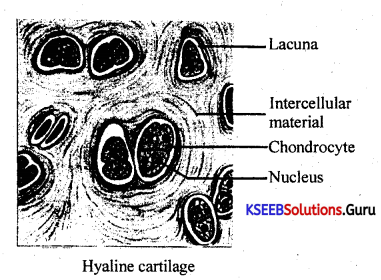
It is a flexible supporting tissue found in-ear pinna and trachea of vertebrates. Cartilage is of two types.
(i) Hyaline cartilage.
(ii) Fibrous cartilage.
![]()
(i) Hyaline cartilage :
1. This cartilage is covered by a fibrous and vascular layer called perichondrium.
2. It is found at the ends of long bones to form joints. It is found in nose, lip, larynx, trachea, etc.
3. Matrix: It is the ground substance composed of glycoproteins. It is also known as chondrin. Matrix is transparent, glossy and free from fibres, hence the name Hyaline cartilage.
4. Chondrocytes: Cells of cartilage are caned chondrocytes or chondroblasts. These are spherical or oval cells with large nucleus at the centre and granular cytoplasm. They secrete the matrix of the cartilage. They get nourishment indirectly from the blood vessels of the perichondrium. Hyaline cartilage gives support and shape to the organs of the body. It also helps in the articulation of bones.
(ii) Fibrocartilage (White fibrous cartilage) :
1. It occurs in pubic symphysis and intervertebral discs.
2. Matrix is made up of glycoproteins secreted by cartilage cells.
However the matrix includes more white fibres, hence the name fibrocartilage or white fibrous cartilage. White fibres of fibro cartilage are made up of collagen and they give strength to the tissue.
4. Fibrocartilage often serves as shock absorber for structures that are subjected to pressure, provides support and helps in fusion. ,
Vascular tissue or Fluid tissue:
Mainly it is a circulatory and transporting tissue. It is of two types, namely:
(a) Blood (b) Lymph.
(a) Blood: It is a fluid tissue consisting of two components.
(a) Blood: It is a fluid tissue consisting of,
1. Plasma.
2. Corpuscles or cells.
1. Plasma:
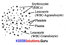
It is a straw yellow (almost colourless) homogenous matrix containing large amount of water (about 90%). A number of gases (O2, CO2) and substances are dissolved in it. Plasma also carries metabolic wastes, hormones, antibodies, etc. Plasma is the matrix and blood corpuscles are suspended in it.
2. Blood corpuscles: They are of 3 types, namely
(i) Erythrocytes or RBCs (Red Blood conpuscles).
(ii) Leucocytes or WBCs (White Blood conpuscles).
(iii) Platelets.
(i) Erythrocytes: They are circular in outline but look like biconcave from sideview. In mammals RBCs are enucleate upon maturation (except those of the family Camellidae). Cytoplasm includes hae¬moglobin, the iron pigment.
Haemoglobin has greater affinity for Or Thus RBCs transport O2 to various tissues. The number of RBCs is about 5 million per cubic mm of blood. They have a short life span of about 120 days.
(ii) Leucocytes: These are large, and colourless cells which are fewer in number. Their number varies from 5000 to 9000 per cubic mm of blood. They are of two types.
a. Granulocytes
b. Agranulocytes.
a. Granulocytes: They are large amoeboid cells with granular cytoplasm. The nucleus shows three to five lobes. They are of three types based on the staining property.
1. Eosinophils
2. Basophils
3. Neutrophills.
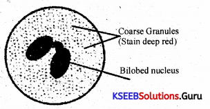
1. Eosinophils: They constitute 2 to 4 % of the leucocytes, i.e., their number ranges from 180 – 360/ mm3 of blood. They are characterised by the presence of coarse cytoplasmic granules that stain deep red with acidic stain like Eosin. The nucleus is bi- lobed. They produce antihistamines that destroy antigen – antibody, and combat against irritants causing allergy.
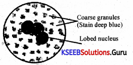
2. Basophils: They constitute only 0.5 – 1 % of the leucocytes i.e., their number range from 45 – 90/mm3 of blood. Basophils are structurally very much similar to eosinophils. However coarse cytoplasmic granules are few in number, and irregular in shape. They stain deep blue with basic stains like methylene blue. They contain heparin, histamine, and serotonin, and are believed to be involved in aller-gic reactions.
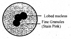
3. Neutrophils: They constitute 60 – 70% of. the leucocytes i.e., their number range from 4.000 – 6.000/mm3 of blood. They are characterised by the presence of fine cytoplasmic granules that stain pinkish with neutral stains. They possess nucleus with 2 -5 lobes. Neutrophils ingest pathogens, hence defensive in function.
![]()
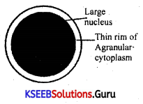
b. Agranulocytes: They are few in number. They are almost oval or spherical in shape with large kidney shaped nucleus. The cytoplasm is smooth or less granulated.
They are of two types.
1. Lymphocytes (produced in lymph nodes).
2. Monocytes (produced in bone marrow).
1. Lymphocytes: They are produced in the lymph nodes (lymphoid tissue) and they constitute 20 – 25% of the leucocytes i.e. their number range from 1800 – 2250/mm3 of blood. They are characterised by relatively large round nucleus surrounded by a thin rim of cytoplasm. They produce antibodies concerned with immunity. Lymphocytes are of two types.
a. T lymphocytes: Concerned with cellular immunity.
b. B lymphocytes: Concerned with humoral immunity.
2. Monocytes: They are produced in the bone marrow (myeloid tissue). They constitute 3 – 8% of the leucocytes i.e .. 270 – 720/mm3 of blood. They have large horse shoe shaped or kidney shaped nucleus. They are phagocytic, and hence destroy invading microor¬ganisms.
Functions of leucocytes :
They defend the body either by ingesting pathogens which are harmful or by producing anti-bodies against foreign bodies.
(iii) Platelets: They are also called thrombocytes. They are small fragments of megakaryo¬cytes with granular form of cytoplasm, Nucleus is absent. The number varies from 2 to 3 lakh per cubic mm of blood. Platelets are brittle, hence break up to release clotting proteins like prothrombin, and thromboplastin to bring about clotting.
General functions of blood:
1. Blood transports food molecules and oxygen to various tissues.
2. Removes CO2 and the nitrogenous wastes from the tissues, and transports them to lungs and kidneys respectively.
3. Maintains constant temperature of the body.
4. Protects the body by producing antibodies and ingesting pathogens.
5. Also carries hormones and vitamins.
Lymph:
It is a colourless mobile fluid or vascular connective tissue which flows in vessels called lymph vessels. Lymph is derived from intersitial or tissue fluid by means of fine, blind and iregular lymph capillaries. It is ultimeately drained into venous blood.
Functions
1. Large Molecules : Plasma proteins and other large macromolecules synthesised by liver and other parts of the body are picked up by lymph for passage to blood.
2. Hormones : Hormones are first passed into lymph before they are carried by blood.
3. Fat Drops : Fat is absorbed from alimentary canal by lymph capillaries called lacteals.
4. Waste Products : A part of the waste products and carbon dioxide are picked up from tissues by lymph to supplement their drainage by blood.
5. Lymphocytes : Lymphocytes are processed by lymph nodes and other lymphatic structures. The same are then passed by lymph into blood.
6. Excess Tissue Fluid : It picks up excess tissue fluid from interstitial spaces.
7. Maintenance of Blood Volume : Lymph is responsible for maintaining blood volume which decreases due to seepage of plasma from blood capillaries into tissue fluid.
8. Pathogens : Pathogens and other foreign particles easily pass into lymph due to the higher permeability of lymph capillaries. They are taken to lymph nodes for destruction.
Organ And Organ System:
An organ is a compact structure made up of one or more types of tissues. For example our heart consists of all the four types of tissues i.e., epithelial, connective, muscular and neural.
An organ along with an accessory organ is called as organ system.
Phereiima posthuma : Earth worm.
• Earthworm belongs to the phylum Annelida, class aligochaeta.
• Habitat: The common Indian earthworm is an important representative of phylum Annelida. It is a burrowing animal. It lives in burrows made in the soft, moist soil, rich in dead and decaying organic matter or humus. It digs its borrows mostly in the soft soils of gardens, lawns or pastures. The burrows may extend vertically downwards upto a depth of 30-60 cms. In spring and summer, the burrows may go deep upto a depth of 180-240 cm where moisture is available. During rainy season, the burrows are flooded and the worms leave the burrows and creep on the moist surface of the soil.
• Habits and Habitat:
1. Earthworm is nocturnal. During the day time, it remains passively in the burrow with its anterior end directed upwards and posterior end downwards. At night, it becomes active. It may come out, of the burrow and roam about. Some times it keeps the posterior end fixed to the burrow and moves the anterior part of the body in a circle. It feeds on soil particles rich in decaying organic matter or humus. It can also ingest directly the bits of leaves and other organic matter. The undigested food is egested as worm casting which appear as heaps on the soil.
2. Earthworm respires through its skin which is very thin and highly vascular. It is always kept moist. Exchange of gases takes place by diffusion through the skin.
3. Locomotion is effected by the contraction and relaxation of muscles of the body wall aided by setae or chaetae. The setae are embedded in setal sacs. They can be protruded or retracted. During locomotion, the anterior part of the body is extended. It is fixed to the substratum by setae. The posterior part of the body is drawn forward towards the anterior part. It is then fixed to the sub-stratum by the setae and again the anterior part is stretched. This process is repeated. Thus, earth worm creeps slowly on the surface of the soil. The setae hold the substratum firmly when the worm is making burrows. The earthworm can move backwards by reversing the direction of setae. The backward movements occur when it has to come out of the burrow. The Earthworm moves at the rate of 25 centimetres per minute.
![]()
4. There are no special sense organs like eyes, ear etc. But the skin of the earthworm has receptor cells, and is highly sensitive to light and soil vibrations. The worm has the sense of smell and can detect food immediately.
5. The Earthworm is hermaphrodite or bisexual or monoecious. It is oviparous. There is no self fertilisation because it is protandrous. Thus, there is cross-fertilisation. During copulation, two earthworms are closely attached with each other by their ventral surfaces in such a way that the head region of one is opposite the tail region of the other. There is mutual exchange of spermatic fluid. Then the two worms separate;
• Features of Pheretima posthuma (Earthworm):
1. Earthworms are metamerically segmented nocturnal animals, live in burrows made in the
soft, moist soil of gardens, lawns, forests, pastures, flower beds and the soil rich in decaying organic matter or humus. They are rarely found in dry, sandy and clayey soils. They usually lie in the upper layers of soils. ‘
2. Earthworm moves by the muscular contraction and expansion of the body, aided by chitinous setae present in the body wail.
3. Its body is elongated, 100 -120 segmented, cylindrical and pointed at both ends. When fully grown, measures about 150 mm in length and about 3 to 5 mm in diameter. The dorsal side of earthworm is more dark brown in colour due to deposition of a brown coloured substance known as porphyrin which is derived out of the’ decayed leaves, on which the earthworm feeds. First segment is called peristomium with an extension of fleshy lobe, prostomium, segments 14 -16 are covered by clitellum.
4. Apertures : Many apertures are found on the external surface which are openings of , coelom, alimentary canal, the excretory and genital organs.
(a) Mouth: It is a crescentriC anterior aperture present in the first segment called peristomium.
(b) Anus: It is a vertical slit-like aperture present at the posterior end of the last segment called anal segment or pygidium.
(c) Female genital pore: It is a single median aperture of the oviducts present on the ventral side of the 14th segment. Eggs pass out of this aperture.
(d) Male genital pores: They are a pair of crescentric openings of the common prostate spermatic ducts present in 18th segment on the ventral side.
(e) Genital papillae: There are two pairs of genital papillae present in the 17th and 19th segments on the ventral side. They bear the minute openings of accessory glands.
(f) Spermatheca pores : They are four pairs of apertures, each pair is present in the grooves of 5/6, 6/7, 7/8 and 89 segments. They are ventrolateral in position.
(g) Nephridiopores: These are minute openings of the integumentary nephridia present in the body walls. They are scattered in all the segments except the first two segments.
(h) Dorsal pores: These are minute pores of coelomic chambers located mid-dorsally, one in each inter- segmental groove, behind the 12th segment.
![]()
5. Body wall: It is covered with a very thick cuticle. The body
wall consists of the following layers,
(a) Cuticle
(b) Epidermis
(c) Circular Muscles
(d) Longitudinal Muscles
(e) Coelomic Epithelium.
Epidermis is one cell in thickness and consists Of,
(i) Mucus secreting cells
(ii) Albumen secreting cells
(iii) Sensory cells
(iv) Supporting cells
(v) Basal cells.
6. The Coelom : There is a true coelom present in earthworm. It is partitioned into so many coelomic compartments with the help of septa. The first septum lies between 4/5 segments and is thin and membranous. In the coelom a milky white fluid known as coelomic fluid is present which is slightly acidic.
There are several types of corpuscles present in the coelomic fluid.
(a) Phagocytes : Amoeboid in shape and they destroy foreign bacteria.
(b) Chloragogen cells : These are yellow coloured, have excretory functions.
(c) Mucocytes : These are fan shaped corpuscles.
(d) Circular nucleated cells.
(e) Eliocytes.
7. Digestive System : The alimentary canal consists of the following parts situated in the segments as mentioned. Mouth is situated on the ventral side of first segment. It opens into the buccal cavity which lies in the first 3 segments. This opens into the pharynx situated in the fourth segment. From 5th to 7th segment is the oesophagus followed by a highly muscular structure known as gizzard situated in the 8th segment. From 9th to 14th segments lies the stomach. From 15th segment till the last segment is the intestine. The intestine is divided into 3 parts known as;
(a) Pretyphiosolar region (15th to 26th segment).
(b) Typhiosolar region (27th to nearly end except last 20-25 segments).
(c) Post-typhiosolar region (situated in the last 20-25 segments).
8. Excretory System: Excretion takes place by means of a large number of coiled tubular structures known as nephridia. These are the excretory units. There are 3 types of nephridia found in earthworm.
(a) Integumentary Nephindia: These are exonephric, present on the inner side of the body wall in all the segments except the first two segments. Their number is 200 to 250 in each segment except in clitellum, where they are 10 times more. These are known as “Forests of nephridia.
(b) Septal Nephridia: These are enteronephric and situated behind the 15th segment attached to the septa, on both sides. Each row has 80 to 100 septal nephredia. They are enteronephric.
(c) Pharyngeal Nephridia: They are enteronephric. Three groups of pharyngeal nephridia are situated in each of the 4th, 5th and 6th segments. Out of all the 3 types of nephridia, only septal nephridia have a funnel-shaped structure known as nephrostome and thus can collect the waste products from the coelomic fluid, other two types don’t have nephrostome.
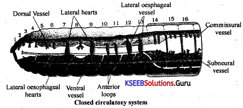
9. Vascular System : It consists of a closed tube of blood vascular system. The haemoglobin is found dissolved in the plasma. It consists of hearts, blood vessels, anterior loops and blood glands.
10. Male Reproductive System : Earthworm is a bisexual (hermaphrodite) animal. The male reproductive organs consists of two pairs of testes situated in 10th and 11th segments. Each testis is enclosed in a fluid filled testis sac. There are 2 pairs of seminal vesicles situated in 11th and 12th segments. The testes sacs communicate with corresponding seminal vesicles.
Just beneath each testes lies a funnel known as spermiducal funnel. From each funnel is given out a duct known as vas deferens. The two pairs of vasa deferentia open into the prostate glands lying from 17th to 20th segments. From each prostate gland is given out a duct known as common prostate spermatic duct. These ducts open on the ventral side of the 18th segment through male genital apertures.
11. Female reproductive organs consist of one pair of ovaries situated in the 13th segment. Just close to each ovary is a funnel, known as oviducal funnel. Both the oviducal funnels open into oviducts. Both the oviducts enter into the 14th segment and open outside through the female genital aperture. Female reproductive organs also include 4 pairs of spermathecae situated in 6th, 7th, 8th, 9th segments. The spermethecae open to the exterior through 4 pairs of spermathecal pores situated in the inter-segmental grooves of 5/6,6/7, 7/8 and 8/9 segments.
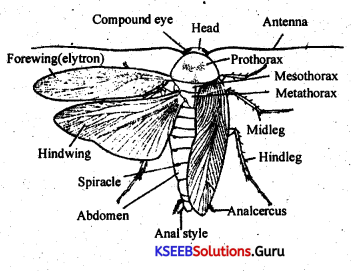
Periplaneta americana (Cockroach):
Introduction:
Cockroach is a hexapod or six legged insect, living in the dwelling places of humans. It is nocturnal active by night and a house hold pest.
Systematic position:
Phylum – Arthropoda.
Class – Insecta (Hexapoda).
Order – Dictyoptera.
Family – Blattidae.
Genus – Periplaneta.
Species – americana.
Habits and Habitat:
Kitchen, store houses, book shelves, radios, televisions, reffigirators, sofa sets and also common in food processing industries like hotels, bakeries besides being common in latrines and sewage canals. It is nocturnal, i.e.-active during nights and remain hiding in dark places during day time.
Symmetry: Body is bilaterally symmetrical. Size and shape: Cockroach may grow to a length of 4 to 5 cms and about 1 cm in breadth.
Exoskeleton: Externally the body is covered by a thick, hard, chitinous cuticle called exoskeleton. Body is segmented, and each segment has chitinous plates called sclerites. The sclerite on the upper side is called tergum, lower side plate is called sternum. These two are 1 laterally connected by pleurites which is a thin membrane. During growth the exoskleton is shed several times and the process is called moulting or ecdysis.
Body structure: Cockroach body is segmented and externally covered by a exoskeleton. The body is divisible into three parts, namely, Head, Thorax and Abdomen.
![]()
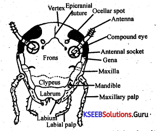
Head: The head is pear shaped and broad on top and narrow down. Wards head is connected to the thorax through a short neck. Words ead has sense organs like antennae, compound eyes and ocelli.
Antennae are long, jointed and movable structures acting as feelers. These are set in antenal sockets.
The compound eyes are large, blackish, and each compound eye is made up of many small functional units called ommatidia.
Mouth parts or trophi of insects in a cockroach:
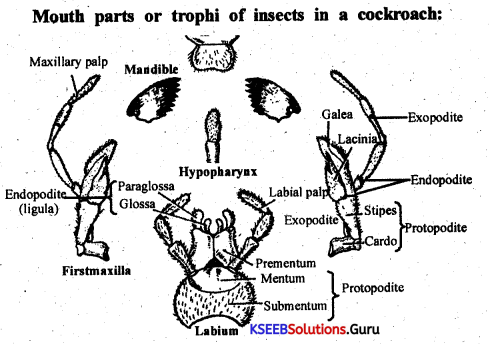
The mouth parts of an insect is called a trophus. In cockroach the trophus includes, labrum, mandibles, first maxilla, second maxilla (or lower lip or labium) and tongue. Mouth parts of cockroach with its mandibles is called mandibulate type or biting and cutting type.
Labrum: It is a thin, mobile and membranous part, it acts as an upper lip.
Mandibles: There are two mandibles. They are lateral to mouth and below the labrum. Their inner edge is toothed and they move from side to side.
First maxilla: These are paired structures, one on each side. The maxilla consists of two basal segments, the cardo and stipes and an upper part with five jointed, movable maxillary palp. At the inner basal end of maxillary palp are two lobes set side by side. They are called laccinia and galea. The lacinia is short, thick and has two stiff bristles. The galea is soft, broader and leaf like.
Second maxilla or labium: It is an unpaired structure. It has two basal segments viz, mentum and submentum. Above mentum are parts like labial palps, glossae and paraglossae. The labial palps are made of three segments and are connected to the sides of mentum through spongy cushion like pads called palpigers. Internal to the labial palps are glossae and paraglossae.
Tongue or hypopharynx: It is tubular and is on the inner side of labium. Most parts connected to feeding are provided with sensory bristles.
Neck: It connects head with the thorax. Neck is slender, soft and supported by cervical sclerites. Muscles of the neck permits movement of head in different directions. On the upper side,the neck is concealed by the expanded prothoracic shield or notum.
Thorax: It is the second division of the body. It consists of three segments. They are prothorax, mesothorax and metathorax. Each thoracic segment is dorsally covered by a chitinous plate called tergum or notum and ventrally by the sternum. These two are laterally connected by a pleuron.
Between the adjacent segments are thin articular membranes which provide flexibility. The tergum of prothorax is broad and projects forward to concael the neck. In the thorax there are three pairs of legs and two pairs of wings.
Thoracic legs: Each segment of thorax has a pair of legs. They are used for walking, running and climbing. Legs’ are jointed structures. Each leg is made up of five segments.
They are coxa, trochanter, femur, tibia and tars.
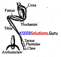
Coxa: Is the first segment, thickest and joins the leg with the thorax.
Trochanter: is the second segment. It is small and is found between coxa and feipur.
Femur: Is the third and the strongest segment of the leg. In front of the femur is the tibia the fourth segment, folowed by tarsus, the fifth division of leg.
Tibia: Is the fourth segment of leg in cockroach and is next to femur. It is slender and beset with many stout spines, the tibial spurs.
![]()
Tarsus: Is formed of five small segments called tarsorneres. The terminal tarsus ends with a pair of curved claws. Between the claws is a soft hollow lobe covered of bristles called pulvillus or arolium.
Wings: There are two pairs of wings in cockroach. First pair is in mesothorax and the second is in metathorax. Wings are by a network of chitinous ridges called veins or nervures.The forewings or the first pair are called wing covers or tegmina (L. tegere = cover). The hindwings are thin, broader, membranous and used in flight These are kept folded at rest and are protected by the first pair of wings.
Abdomen: It is the third and largest division of the body. In the adult, it consists of ten segments and eleven in the embryonic stages. The first seven segments are usually distinct. The eighth and ninth are tucked in and are concaeled by the over growth of seventh tergum.
There is a pair of short eighteen segmented anal cerci, one On either side of tenth segment serving sensory function. In male cockroaches there are a pair of needle like, unjointed structures projecting ninth segment called as anal styles. They occur between anal cerci.
The sexes are distinct. Males have genital opening between tenth and niftth segments. In females the genital opening is located ventrally in the eighth segment. The genital openings in both sexes are surrounded by gonapophyses. In females immediately behind the genital opening is the genital pouch or gynatrium, a chamber formed by intucking of eighth and ninth segments. In males genital pouch is rudimentary or reduced.
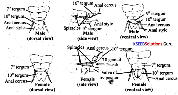
Female Cockroach:
1. Abdomen broad and short.
2. Anal styles absent.
3. A brood pouch enclosed in the 7th, 8th and 9th sterna is present
4. Antennae shorter.
5. Seventh abdominal sterna are visible only.
6. Only the first seven abdominal sterna are visible (only).
7. Genital aperture lies on the 8th sternum.
8. Gonapophyses consists of pairs of plates.
Male Cockroach:
1. Abdomen narrow and long.
2. Anal styles present. They are in the form of unjointed appendages attached to the 9the segment.
3. No brood pouch.
4. Antennae longer.
5. Seventh abdominal tergum covers only the 8th sternum.
6. All the nine abdominal sterna are visible.
7. Gential aperture lies below the anus between the 10th tergum and 9th sternum.
8. Gonapophyes consists of 3 phallomeres only.
Salivary gland:
On either side of oesophagus and crop there are paired salivary glands. It consists of paired salivary receptacles and from each receptacle arises a duct called salivary receptacular duct. Ducts of both receptacles are united to form a common receptacular duct. Around each receptace there are granular leaf like structures called salivary glands. From salivary glands, a duct called salivary duct arises. The salivary ducts of both the glands are united for form a common salivary duct.
Both the common receptacular duct and salivary duct are united to form a common delivery receptacular duct or efferent duct. This duct ends at the tongue called hypopharynx. The salivary glands secrete a salivary juice which is stored in receptacles. The salivary juice contains an enzyme ptyalin, which digests carbohydrates.
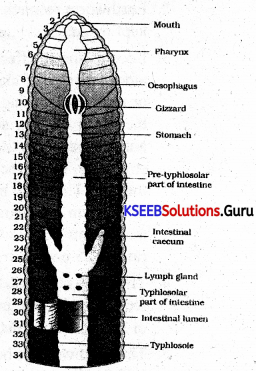
The foregut is the begining part of the digestive canal. It includes mouth, buccal cavity, oesophagus and gizzard. Associated with the foregut, there are well developed salivary glands. The buccal cavity leads into a narrow tubular oesophagus. The oesophagus connects to a thin sac called crop followed by a thick walled pot-shaped gizzard (or proventriculus). Inside gizzard are six chitinous cuticular teeth.
In the gizzard food is subjected to grinding, mixing and filtering processes. Hence the gizzard is often called as grindmill of insect. Food is mixed with enzymes of hepatic caecae. The bristles present in the lower part of the gizzard act as filtering agents and push up larger particles to be crushed again and allow (filter) only minute particles to enter the mesenteron.
The mesenteron or midgut is more a tubular structure. It is lined by endoderm. At the junction of stomodeum and mesentron are six to eight finger like blind diverticulae called hepatic caecae. The digested food is absorbed in the mesentron.
The hindgut is long and coiled. This is the final division and is made up of narrow ileum, a wider colon and sac like rectum. The midgut and hindgut are marked out clearly by the presence of slender, hair like, yellow coloured, fragile, malpighian tubules. Rectum communicates outside through anus.
• Circulatory System is of open type. The blood is colourless, without haemoglobin, known as haemolympl. The body cavity (Haemocoel) is divided into 3 cavities by diaphragms known as pericardial sinus, peri viscera and perineural sinuses.
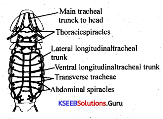
Respiratory system:
(i) Heart is situated in the pericardial sinus, and is made up of 13 segmentally arranged funnel shaped chambers. There are fan-shaped muscles attached to the heart in each segment known a salary muscles, which help in opening and closing of the ostia. The haemolymph flows in the heart in the forward direction.
(ii) Excretion is carried out by about 60 fine, yellow, unbranched, thread-like blind tubes called malphigian tubules. The main excretory product is uric acid. Respiration is carried out through trachea. Compound eyes are formed of a number of optical units called ommatidia and the image formed is mosaic.
Nervous System:
Higher animals have nervous system which control various activities of the body. Generally, this system includes the brain, nerve cord and nerve fibres. In invertebrates the brain is situated dorsally in the head and nerve cords ventrally in the body. The nervous system in cockroach is built on the same plan as that found in annelids. However, the number of ganglia do not correspond with the number, of segments. Thus reduction in the number of ganglia, development of special sense organs, locomotar respiratory organs, etc is an important evolutionary change. The nervous system of Cockroach includes central, peripheraland sympathetic systems.
![]()
Central nervous system :
A pair of ganglia are situated below the oesophagus and are called as sub oesophageal ganglia. The supra and sub oesophageal ganglia are laterally connected by circum oesophageal connectives forming a nerve ring.
Nerve cords:
There are two nerve cords extending from the back of sub-oesophageal ganglia. These are found on the mid ventral region of the body and are connected by nine pairs of ganglia (three in the thorax and six in the abdomen). The last or the sixth pair of abdominal ganglia is larger than the rest.
The brain (supra-oesophageal ganglia) sends out nerves to the compound eyes and antennae. The sub-oesophageal ganglia, similarly supply nerves to many of the mouth parts.
In the thoracic region, the thoracic ganglia supply nerves to legs, wings, trachea, chambers of heart and other structures of the corresponding segments. The abdominal ganglia supply nerves to various parts of that region. The last pair of ganglia innervates the sixth, seventh, eighth, ninth and tenth abdominal segments and send branches to anal cerci and anal styles.
Peripheral nervous system: Nerves arising from the brain and ganglia of the nerve cords reach various organs. Such nerves in the bddy represent the peripheral nervous system.
Sympathetic nervous system: This system is comprised of the frontal ganglia in front of the brain, another smaller pair behind brain and a large ganglia on the dorsal side of crop, together, with their connectives joining all of them to the brain. Nerves arising from these reach muscles, viscera (alimentary canal), spiracles and control their activities.
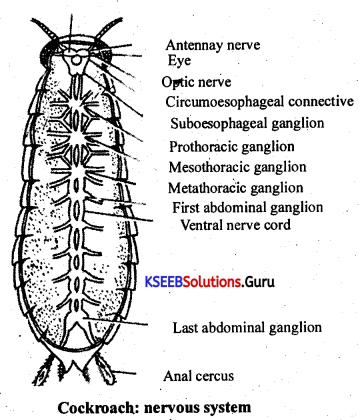
• Male reproductive organs. It consists of one pair of testes situated from 3rd to 6th abdominal segments. Each testes gives out a vas deferens. Both the vasa deferentia run posteriorly and open into an ejaculatory duct, where a gland known as utricular gland is situated. This gland has 3 types of tubules known as long peripheral tubules, short central tubules and one pair of seminal vesicles which store the sperms. Ejaculatory duct runs posteriorly and opens to the exterion through an aperture known as male gonopore.
• Around the male gonopore, are situated a few chitinous structures known as male gonapophysis. They are is made up of 3 phallomeres-left, right and ventral phallomeres. The left phallomere has structures known as Titilator, Pseudopenis and Asperate lobe. All these help during copulation. The sperms of cockroach are released in the form of a bundle known as spermatophore which is surrounded by 3 layers.
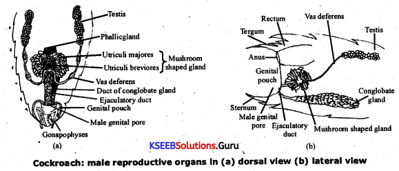
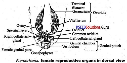
• Female Repreductive organs. It consists of one pair of ovaries lying from 3rd to 6th abdominal segments. Each ovary is made up of 8 beaded structures known as ovarioles, in which the eggs are arranged in a linear fashion in the order of development. From each ovary arises a duct known as oviduct. Both the oviducts join to make a common duct known as vagina which opens into the genital chamber through the female gonopore. In the gonopore lie a pair of spermathecae, one pair of collateral glands and six chitinous plates known as ovipositor.
• Ootheca formation and egg laying. The eggs are laid in a brown, horny purse-like egg case called ootheca. The egg- cases are laid in a dark place in a crevice, in dirt, pushed down by the ovipositor. Each egg-case contains 16 fertilized ova (eggs) in two vertical rows. Each egg in the egg case hatches into a colourless young cockroach called nymph.
• Nymph hatched undergoes gradual metamorphosis (6- 9 moults) to form into an adult.
Rana Tigrina (Frog):
Habitat of frog :
Rana tigrina is commonly found in or near water bodies like ponds, lakes, ditches, wells etc. It is an amphibious animal.
Systematic position
Phylum – Chordata.
Sub phylum – Vertebrata or Craniata.
Class – Amphibia.
Order – Anura.
Genus – Rana.
Species – tigrina.
• Their body temperature varies with the temperature of the environment, and such a called cold blooded animals or poikilotherms.
• Frogs are not seen during peak summer and winter. During this period they take shelter in deep burrows to protect them from extreme heat and cold. This is called as summer sleep (aestivation) and winter sleep (hibernation).
Morphology :
• The skin is smooth and slippery due to the presence of mucus. The skin is always maintained in a moist condition.
• The colour of dorsal side of body is generally olive green with dark irregular spots. On the ventral side the skin is uniformly pale yellow.
• Body of a frog is divisible into head and trunk. Neck and tail are absent. Above the mouth, a pair of nostrils are present. Eyes are bulged and covered by a nictitating membrane that protects them while in water. On either side of the eyes, a membranous tympanum (ear) receives sound signals. The forelimbs and hind limbs help in swimming, walking, leaping and burrowing. The hind limbs end in five digits and they are larger and muscular than the fore limbs that end in four digits. Feet have webbed digits that help in swimming.
• Male frogs can be distinguished by the presence of sound producing vocal sacs and also a copulatory pad on the first digit of the fore limbs which are absent in female frogs.
Anatomy:
• The body cavity of frogs accommodates different organ systems such as digestive, circulatory, respiratory, nervous, excretory and reproductive systems, with well developed structures and functions.
![]()
• The digestive system consists of alimentary canal and digestive glands. The alimentary canal is short because frogs are carnivores and hence the length of intestine is reduced. The mouth opens into the buccal cavity that leads to the oesophagus through pharynx.
• Oesophagus is a short tube that opens into the stomach which in turn continues as the intestine, rectum and finally opens outside by the cloaca;
• Liver secretes bile that is stored in the gall bladder.
• Pancreas, a digestive gland produces pancreatic juice containing digestive enzymes.
• Digestion of food takes place by the action of HCI and gastric juices secreted from the walls of the stomach. Partially digested food called chyme is passed from stomach to the first part of the intestine, the duodenum. The duodenum receives bile from gall bladder and pancreatic juices from the pancreas through a common bile duct.
Bile emulsifies fat and pancreatic juices digest carbohydrates and proteins. Final digestion takes place in the intestine. Digested food in the inner wall of intestine called villi and microvilli. The undigested solid waste moves into, the rectum and passes out through cloaca.
• Respiratoy System: Frogs respire on land and in the water by two different methods. In water, their skin acts as the aquatic respiratory organ, and it is exchanged through the skin by diffusion..
• On land, the buccal cavity, skin and lungs act as the respiratory organs. The respiration by lungs is called pulmonary respiration. The lungs are a pair of elongated, pink coloured sac-like structures present in the upper part of the trunk region (thorax). Air enters through the nostrils into the buccal cavity and then to lungs. During aestivation and hibernation gaseous exchanges takes place through the skin.
• The vascular system of frog is a well-developed closed type. Frogs have a lymphatic system also. The blood vascular systeminvolves heart, blood vessels and blood. The lymphatic system consists of lymph, channels and lymph nodes.
• Their heart is a muscular structure, situated in the upper part of the body cavity. It has three chambers, two atria and one ventricle and is covered by a membrane called pericardium. A triangular structure called sinus venosus joins the right atrium. It receives blood through the major veins called vena cava.
The ventricle opens into a sac like conusarteriousus on the ventral side of the heart. The blood from the heart is carried to all parts of the body by the arteries (arterial system). The veins collect blood from different parts of body to the heart and form the venous system.
• Excretory system : The excretory system consists of a pair of kidneys, ureters, cloaca and urinary bladder. These are compact, dark red and bean like structures situated a little posteriorly in the body cavity on both sides of the vertebral column. Each kidney is composed of several structural and functional units called uriniferous tubules or nephrons. Two ureters emerge from the kidneys in the male frogs.
The ureters act as urinogenital duct and oviduct opens seperately in the cloaca. The thin-walled urinary bladder is present ventral to the rectum, which also opens in the cloaca. The frog excretes urea and thus is a ureotelic animal. Excretory wastes are carried by blood into the kidney where it is separated and excreted.
• Nervous system : The system for control and coordination is highly evolved in the frog. It includes both a neural system and endocrine glands.
• The prominent endocrine glands found in frog are pituitary, thyroid, parathyroid, thymus, pineal body, pancreatic islets, adrenals and gonads.
• The nervous system is organised into a central nervous system (brain and spinal cord), a peripheral nervous system (cranial and spinal nerves) and an autonomic nervous system (sympathetic and parasympathetic). There are ten pairs of cranial nerves arising from the brain. Brain is enclosed in a bony structure called brain box (cranium). The brain is divided into forebrain, midbrain and hindbrain.
Forebrain includes olfactory lobes, paired cerebral hemispheres and unpaired diencephalon. The midbrain is characterised by a pair of optic lobes. Hindbrain consists of cerebellum and passes out through the foramen magnum and continues as the spinal cord, which is enclosed in the vertebral column.
• Frog has different types of sense organs, namely organs of touch (sensory papillae), taste (taste buds), smell (nasal epithelium), vision (eyes) and hearing (tympanum with internal ears). Out of these, eyes and interal ears are well-organised structures and the rest are cellular aggregations around nerve endings.
• External ear is absent in frogs and only tympanum is present.
• Reproduction system : Male reproductive organs consist of a pair of yellowish ovoid testes, which are found adhered to the upper part of kidneys by a double fold of peritoneum called mesorchium. Vasa efferentia are 10-12 in number that arise from testes. They enter the kidneys on their side and open into Bidder’s canal. Finally it communicates with the urinogenital duct that comes out of the kidneys and opens into the cloaca. The cloaca is a small, median chamber that is used to pass faecal matter, urine and sperms to the exterior.
• The female reproductive organs include a pair of ovaries. The ovaries are situated near kidneys and there is no functional connection with kidneys. A pair of oviduct arising from the ovaries opens into the cloaca separately. A mature female can lay 2500 to 3000 ova at a time.
• Fertilisation is external and takes place in water. Development involves a larval stage called tadpole. Tadpole undergoes metamorphosis to form the adult.