2nd PUC Biology Notes Chapter 11 Biotechnology: Principles and Processes

Karnataka 2nd PUC Biology Notes Chapter 11 Biotechnology: Principles and Processes

→ Biotechnology is a synthetic co-integrated branch of science which utilises the knowledge of biochemistry, chemical engineering and microbes, in tapping the inactive behaviour of microbes and cultured cells for technical applications.

→ It can be defined as a technology in which microorganisms and isolated cells, tissues and organs of desired plants are employed to produce novel varieties of desired products for the welfare of mankind. The term biotechnology was coined by “Karl Erecky”.

2nd PUC Biology Notes Chapter 11 Biotechnology: Principles and Processes

Principles of Biotechnology:

I. Genetic engineering or Genetic surgery:
The method of isolating or synthesizing the desired gene and its insertion in the genome of the other organisms to produce hybrid DNA or recombinant DNA is called genetic engineering.
II. Maintenance of sterile environment

Tools used in genetic engineering are as follows.

  1. Desired gene
  2. Vector or Vehicle DNA
  3. Enzymes.
  4. Host cell
  5. Bioreactors.

1. Desired gene: The gene of interest which is also called desired gene or foreign DNA, has to be obtained from its source or is to be synthesized artificially in the lab.

2. Vectors: These are molecular vehicles which carry the desired gene from one organism to the other organism. There are different types of vectors namely plasmids, bacteriophages, cosmids, phasmids etc [cosmid means plasmid + cohesive ends of phage DNA, Pasmid means phage DNA+a small part of plasmid]

Plasmids:
The small circular and extra chromosomal double stranded DNA molecules present in the cytoplasm of bacteria are called plasmids. They show self replication. They contain 2 to 8 thousands of nitrogen base pairs. They also contain some genes, like antibiotic resistant genes and genes for expression of some characters. They can be cut at specific sites using restriction enzymes and desired genes can be inserted into these plasmids. There are different kinds of plasmids, e.g. F-plasmid [Sex, plasmid], R-plasmid [resistant plasmid], col plasmid [colicins-that kill some strains]. In additions to these there are some genetically engineered plasmids like pBR-322 [Plasmid of Boliver and Rodriguez] and pUC-18 [Plasmid of University of California]
2nd PUC Biology Notes Chapter 11 Biotechnology Principles and Processes 1

2nd PUC Biology Notes Chapter 11 Biotechnology: Principles and Processes

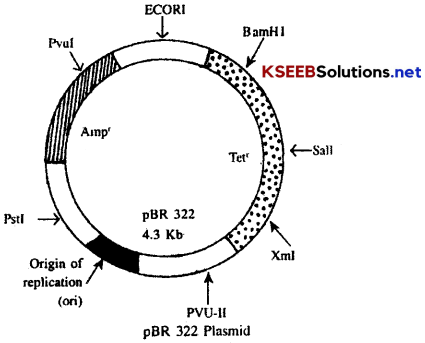
Structure of pBR-322:
pBR-322 is a natural plasmid and F+ plasmid. It is about 4.3 kb in size. It is a plasmid with an ori site (origin of replication), two antibiotic resistance sites, selectable markers for Amphicillin resistance (Ampr) and Tetracycline resistance (Tetr). It has thirteen unique sites in different regions out of which seven are important. A unique site is a specific restriction enzyme (REN) recognition site called ECORI.

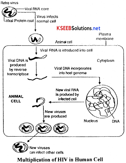
Vectors for cloning genes in plants and animals are Ti plasmid isolated from Agrobacterium tumefaciens in plants and retrovirus are now made non pathogenic and are used to deliver gene into animal cells.

Note: Insertional inactivation: When a gene or recombinant DNA is inserted within the coding sequence of a vector, the coding sequence responsible for an enzyme or a particular character becomes inactivated. This is known as Insertional inactivation.

3. Enzymes: There are two main types of enzymes that are mainly used in the genetic engineering. They are RENs and Lygases.

(a) Restriction endonucleases-REN: The nuclear enzymes that break DNA at specific sites are called restriction endonucleases [REN], The RENs are popularly known as Biomolecular scissors. These are first discovered by Hamilton smith. There are a number of restriction endonucleases. Each enzyme recognizes specific sequence of DNA and cuts both the strands of DNA. For example, Eco RI recognizes the base sequences in DNA. REN may cut both the strands of DNA at the same position or at different positions. [When both the strands of DNA molecule are cut at the same position and no nucleotides are left unpaired, it results in blunt ends] [Symmetric ends] [When each strand of DNA molecule is cut at a different position, it results in sticky ends] [Asymmetric ends] e.g:- Eco RI, Hind III, Sal I, Bam 1.

Note: Restriction enzymes belong to a larger class of enzymes called nucleases. These are of two kinds : exonucleases and endonucleases. Exonucleases remove nucleotides from the ends of the DNA whereas, endonucleases make cuts at specific positions within the DNA.

(b) DNA Lygase:- The enzyme that joins the cut ends of DNA fragments is called DNA lygase. It is popularly called biomolecular stitchers [Molecular glues]. The enzyme joins either the blunt ends or the sticky ends of DNA.

4. Separation and isolation of DNA fragments:
The cutting of DNA by restriction endonucleases results in the fragments of DNA. These fragments can be separated by a technique known as gel electrophoresis. Since DNA fragments are negatively charged molecules they can be separated by forcing them to move towards the anode under an electric field through a medium/matrix. Nowadays the most commonly used matrix is agarose which is a natural polymer extracted from sea weeds. The DNA fragments separate (resolve) according to their size through sieving effect provided by the agarose gel. Hence, the smaller the fragment size, the farther it moves.

The separated DNA fragments can be visualised only after staining the DNA with a compound known as ethidium bromide followed by exposure to UV radiation (you cannot see pure DNA fragments in the visible light and without staining). You can see bright orange coloured bands of DNA in a ethidium bromide stained gel exposed to UV light. The separated bands of DNA are cut out from the agarose gel and extracted from the gel piece. This step is known as elution. The DNA fragments purified in this way are used in constructing recombinant DNA by joining them with cloning vectors.
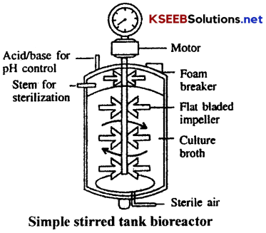
5. Bioreactor:
It is a vessel designed to carry out biological reactions under controlled conditions. It is useful to regulate the contact between the catalyst and its substrate. Commercial bioreactors are generally used to produce the products on a large scale. It is made up of a stainless steel vessel or tank with some controlling units for PH, temperature and nutrient concentrations. It is also provided with pressure gauge inlets for nutrients and filtered air. It is also provided with a coil of pipe for letting in cold water or steam.
2nd PUC Biology Notes Chapter 11 Biotechnology Principles and Processes 2

Uses of bioreactors:

  • They are used to produce primary metabolites like ethanol, acetone, butanol, lysine, glutamic acid etc.
  • They are used in the production of monoclonal antibodies, vaccines etc.
  • They are used in tissue culture.
  • They are used in shoot and root culture of some plants.

2nd PUC Biology Notes Chapter 11 Biotechnology: Principles and Processes

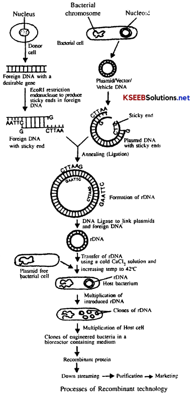
Process of Genetic Engineering:

(a) Isolation of genetic material (DNA): In recombinant DNA technology, it is essential to isolate DNA in pure form free from other macro molecules. Since DNA molecule is enclosed with the membrane in the cell. we have to break open the cell to release DNA along with other macromolecules like RNA, proteins, polysaccharides and lipids. This is carried out in bacterial cells, plant and animal cells with certain enzymes.

The other macro molecules can be removed by appropriate treatment with specific enzymes. Finally, the purified DNA molecules are precipiatated out after the addition of chilled ethanol and this can be seen as collection of fine threads in the suspension.

(b) Cutting of DNA at specific locations: The isolated purified DNA molecule is cut (cleaved) with the help of a suitable enzyme called restriction endonuclease, into segments with sticky ends.

(c) Gel electrophoresis: The cut DNA fragments are separated by gel electrophoresis using agarose gel. DNA is a negatively charged molecule, hence it moves towards the positive electrode (anode).

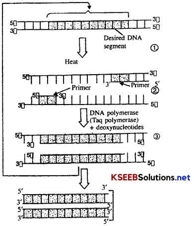
(d) Amplification of Gene of interest using PCR: Amplification of gene is ‘a process of making many copies of a gene’. It is achieved by using a technique called Polymerase Chain Reaction (PCR).

Requirements: 1
→ A DNA containing the desired segment to be amplified

→ Two nucleotide primers (about 20 base long) specific i.e. complementary to the two 3 – ends of the desired segment.

→ The four deoxynculeoside triphosphate i.e. TPP (Thymidine triphosphate) dCTP (Deoxycytidine triphosphate) dATP (deoxyadenosine triphosphate) and dGTP (deoxyguanosine triphosphate)

→ A thermostable DNA polymerase i.e., Taq DNA polymerase.
2nd PUC Biology Notes Chapter 11 Biotechnology Principles and Processes 3

2nd PUC Biology Notes Chapter 11 Biotechnology: Principles and Processes

Procedure of PCR:
1. The DNA from the desired segment to be amplified, an excess of the two primer molecules, the four deoxyribose triphosphates and DNA polymerase are mixed together in a reaction mixture in a eppendorf tube with sufficient quantities of Mg++. The eppendorf tube is placed in the PCR unit and the following operations are performed sequentially.

2. Denaturation: The reaction mixture is first subjected to a temperature between 90-98°C (commonly 94°C). It results in the separation of two strands of DNA due to the breakage of hydrogen bonds. This is called denaturation. Each strand of DNA acts as a template strand for DNA synthesis. The duration of this step in the first cycle of PCR is usually 2 minutes at 94°C.

3. Annealing (anneal=join): The mixture is now cooled to a low temperature (40-60°C). During this step, two oligonucleotide primers, complementary to a region of DNA, anneal (hybridize) one to each 3 end of DNA strand. The duration of annealing step is usually one minute during the first as well as the subsequent cycles of PCR.

4. Primer extension: During this step, the enzyme taq DNA polymerase extend the primers using nucleotides and DNA templates. The two primers extent towards each other in order to get two new strands of DNA (at 5) end. The duration of primer extension is usually 2 minutes at 72°C. The amplified fragment, if required can now be used to ligate with vector for further cloning. The taq DNA polymerase remains active, during the high temperature induced denaturation of double stranded DNA.

(e) Insertion of recombinant DNA into the host: (competent host)
1. Electroporation: The bacterial cell is placed in a solution with cold CaCl2 solution followed by placing them at 42°C intermittently. This results in the development of pores in the cellmembrane. Now the recombinant plasmid migrates into the host cell and the bacterial cell gets transformed.

2. Microinjection : It is direct injection of a desired gene into the nucleus of an animal cell by micro syringe.

3. Biolistics : Here a suitable plant-cell is bombarded with high velocity microparticles (l-2nm) of gold or tungsten coated with DNA inorder to introduce DNA into the cell.

(f) Obtaining the Foreign Gene Product:
→ The transgene expresses itself in the form of protein (s) under appropriate conditions.

→ The product (s) can be extracted from the medium by employing a suitable procedure.

→ The transgenic cells may be cultured in the laboratory to obtain the transgene product on a small scale.

→ The transgenic cells can also be cultured/multiplied in a continuous culture system, in which the used medium is drained out from one side and fresh medium is added from the other side for the production of larger biomass and the desired product.

→ Bioreactors are used for processing large volumes of culture for obtaining the product of interest in sufficient quantities on a commercial scale.
2nd PUC Biology Notes Chapter 11 Biotechnology Principles and Processes 4

2nd PUC Biology Notes Chapter 11 Biotechnology: Principles and Processes

(g) Downstream Processing: The products formed in a bioreactor have to be subjected through a series of processes before they are ready for marketing as finished products. The various processes used for the recovery of useful products are collectively called downstream processing. The processes include separation and purification of the product, addition of suitable preservatives and a stringent quality control testing etc. Such formulation has to undergo strict trials as in the case of drugs. These quality control testing and clinical trials vary from product to product.

2nd PUC Biology Notes

2nd PUC Biology Notes Chapter 10 Microbes in Human Welfare

Karnataka 2nd PUC Biology Notes Chapter 10 Microbes in Human Welfare

→ Microbes are present everywhere in soil water, air, inside our bodies and that of other animals and plants.

→ Microbes belong to diverse groups of organisms protozoa, bacteria, fungi, microscopic plants, viruses, viroids, and also prions.

Microbes in Household Products:

→ Microbes have been used in different ways by mankind from time immemorial in the preparation of household products. Some of the household products obtained from microbial activity are:

→ Dairy Products: A number of bacteria like Lactobacillus and some others commonly called Lactic Acid Bacteria (LAB) are used in the preparation of several dairy products.

2nd PUC Biology Notes Chapter 10 Microbes in Human Welfare

Lactic acid fermentation:

Lactic acid causes coagulation of milk protein casein. Milk is converted into products like curd, yoghurt and cheese.
1. Curd: For making curd, milk is boiled and then the temperature of milk is brought to about 40°C by keeping it in a cool place. Now a small amount of curd (the innoculum or starter containing Lactobacillus acidophilus) is added to the milk. The fermentation process begins which converts milk sugar lactose into lactic acid. The acid coagulates milk protein by partially digesting it so that a thick concentrated curd is prepared. Curd is more nutritious than milk as it contains increased quantity of vitamin B12.

2. Yoghurt (= Yogurt): It is prepared by curdling milk with the help of Streptococcus thermophilus and Lactobacillus bulgaricans. The temperature is maintained at about 45°C for four hours.

3. Butter Milk: The acidulated liquid left after churning out butter from curd is butter milk.

4. Cheese: It is a protein rich nutritive preparation obtained after fermentation and curdling of milk. It consists of milk curd that has separated from liquid part or whey. Depending upon the water content present in milk / curd, cheese is of three types.

  • Raw cheese,
  • Unripened cheese (cottage cheese), and
  • Ripened cheese.

→ Bread: A small amount of baker’s yeast (Saccharomyces cerevisiae) is added to wheat flour at the time of its kneading. The kneaded flour or dough is kept for a few hours in a warm place. It results in swelling of the dough called leavening. The leavening is caused by the actions of three types of enzymes secreted by the yeast. These are amylase, maltase and zymase.

→ Microbes in Industrial Products: A number of industrial products valuable to human beings are produced with the help of microbes. The two common ones are alcoholic beverages and antibiotics.

→ Alcoholic Fermentation: Microbes especially yeasts have been used for the production of alcoholic beverages from time immemorial. Yeast species used in alcoholic fermentation are Saccharomyces cerevisiae (Brewer’s yeast). S. ellipsoides (wine yeast), S. cake (sake yeast) and S. pireform is (Ginger Beer / Ale yeast).

→ Substrates containing starch juice from sugar cane or molasses or sugar beet are the common substrates for the production of ethanol. Ethanol is produced by continuous fermentation. The process is carried out in large fermenters. Fermentation is carried out at pH5, at a temperature of 35°C, but the cultures and culture conditions are different. Varieties of alcoholic drinks are made from the same microorganism using different substrates. Starch from barley grains is used in the preparation of beer. Wine is produced from the sugar in grapes.

2nd PUC Biology Notes Chapter 10 Microbes in Human Welfare

Note: After fermentation is over, the cells are separated to get a biomass of yeast cells which are used as SCP (single Cell Protein) for animal feed.

→ Depending upon the raw material and the type of processing, the alcoholic drinks are of two types:

1. Without Distillation: Wine and beer are produced without distillation. Beer is an undistilled product of grain mash fermentation, while wine is produced from fruit juice without distillation.

2. With Distillation: Whisky, brandy and rum are produced by distillation of the fermented broth.

Antibiotics:

(a) Antibiotics are chemical substances, which are produced by some microbes and can kill or retard the growth of other (disease causing) microbes.

(b) The term antibiotics was coined by Waksman( 1942).

(c) An antibiotic may be broad spectrum i.e., it can kill or destory a number of pathogens that belong to different groups with different structure and wall composition, or specific i.e., effective against only one type of pathogen.

(d) Most of them are produced by actinomycetes, specially the genus Streptomyces and filamentous fungi.

(e) Production of Antibiotics: For commerical production of antibiotics, the sterilized nutrient medium is inoculated with suitable strain of the micro-organism. The nutrient medium is provided with optimum pH, aeration, temperature, antifoaming agent and antibiotic pre-cursor. The microorganism grows and secretes antibiotic in the medium. When sufficient antibiotic has diffused into the medium, the microorganism is separated and the antibiotic is extracted from the medium by precipitation, absorption or solvent. It is purified, concentrated and bioassayed before packing.

Chemicals, Enzymes and other Bioactive Molecules:

1. Aspergillus niger (a fungus) is used in the production of citric acid, Acetobacter aceti (a bacterium) for acetic acid; Clostridium butylicum (a bacterium) for butyric acid and Lactobacillus (a bacterium) for lactic acid.

2. Yeast (Saccharomyces cerevisiae) is used for the commercial production of ethanol.

3. Microbes are also used for production of enzymes. Lipases are used in detergent formulations and are helpful in removing oily stains from the laundry.

Bottled juices are clarified by the use of pectinases and proteases. Streptokinase produced by the bacterium streptococcus and modified by genetic engineering is used as a ‘clot buster’ for removing clots from the blood vessels of patients who have undergone myocardial infaction leading to heart attack.

4. Another bioactive molecule, cyclosporin A, that is used as an immunosuppressive agent in organ – transplant patients, is produced by the fungus Trichoderma polysporum.

5. Statins produced by the yeast Monascus pupureus have been commercialised as blood – cholesterol lowering agents. They act by competitively inhibiting the enzyme responsible for synthesis of cholesterol.

Microbes In Sewage Treatment: Sewage refers to the municipal waste water generated in cities and towns that contains human and animal excreta and other domestic wastes. Large quantities of waste water are generated everyday in cities and towns. Sewage contains large amount of organic matter and microbes. Many of the microbes present in sewages are pathogenic. Therefore, the sewage cannot be discharged into natural water bodies like rivers and streams directly.

To make the sewage less polluting, it has to be treated in Sewage Treatment Plants (STPs). Treatment of waste water is done by the heterotrophic microbes naturally present in the sewage. This treatment is carried out in two stages: Primary treatment and secondary treatment. The waste water can be passed into rivers after secondary treatment. In some cases, tertiary treatment is also carried Out which removes nutrients by chemical process.

2nd PUC Biology Notes Chapter 10 Microbes in Human Welfare

Primary Treatment:
This step involves physical removal of floating and suspended solids from sewage through filtration and sedimentation. Initially, floating debris is removed through sequential fi Iteration. The filtrate is kept in large open settling tanks where grit (sand, silt and small pebbles) are removed by sedimentation. Sometimes, alum or iron sulphate is added for flocculation and settling down of solids. The sediment is called primary sludge, while the supernatant is called effluent. The primary sludge is subjected to composting or land fill. The effluent from the primary settling tank is taken for secondary treatment.

Secondary treatment or Biological treatment:
The primary effluent is passed into large aeration tanks where it is constantly agitated mechanically and air is pumped into it. This allows vigorous growth of useful aerobic microbes into floes (masses of bacteria associated with fungal filaments to form mesh like structures). While growing, these microbes consume the major part of the organic matter in the effluent. This significantly reduces the BOD (biochemical oxygen demand) of the effluent.

BOD refers to the amount of the oxygen that would be consumed if all the organic matter in one litre of water were oxidised by bacteria. The sewage water is treated till the BOD is reduced. The BOD test measures the rate of uptake of oxygen by micro-organisms in a sample of water and thus, indirectly. BOD is a measure of the organic matter present in the water. The greater the BOD of waste water, more is its polluting potential.

Once the BOD of sewage or waste water is reduced significantly, the effluent is then passed into a settling tank where the bacterial ‘floes’ are allowed to sediment. This sediment is called activated sludge. A small part of the activated sludge is pumped back into the aeration tank to serve as the incolulum. The remaining major part of the sludge is pumped into large tanks called anaerobic sludge digesters. Here, other kinds of bacteria, which grow anaerobically, digest the bacteria and the fungi in the sludge. During this digestion, bacteria produce a mixture of gases such as methane, hydrogen sulphide and carbon dioxide. These gases form biogas and can be used as a source of energy as it is inflammable.

The effluent from the secondary treatment plant is generally released into natural water bodies like rivers and streams.

Note River action plan: The ministry of environment and forests has initiated Ganga Action plan and Yamuna Action Plan to save these major rivers of our country from pollution. Under these plans, it is proposed to build a large number of sewage treatment plants so that only treated sewage may be discharged in the rivers.

Microbes in Production of Biogas:

Biogas is a mixture of gases (containting predominantly methane) produced by the microbial activity and which may be used as fuel. Microbes produce different types of gaseous end-products during growth and metabolism. The type of the gas produced depends upon the microbes and the organic substrates they utilise.

Bacteria, which grow anaerobically on ceilulosic material, produce large amount of methane along with CO2 and H2 These bacteria are collectively called methanogens, and one such common bacterium is methanobacterium. These bacteria are commonly found in the anaerobic sludge during sewage treatment. These bacteria are also present in the rumen (a part of stomach) of cattle. A lot cellulosic material present in the food of cattle is also present in the rumen.
2nd PUC Biology Notes Chapter 10 Microbes in Human Welfare 1

Note: In rumen, these bacteria help in the breakdown of cellulose and play an important role in the nutrition of cattle.

2nd PUC Biology Notes Chapter 10 Microbes in Human Welfare

The excreta (dung) of cattle, commonly called gobar, is rich in these bacteria. Dung can be used for generation of biogas, commonly called gobar gas.

The biogas plant consists of a concrete tank (10-15 feet deep) in which bio-wastes are collected and a slurry of dung is fed. A floating cover is placed over the slurry, which keeps on rising as the gas is produced in the tank due to the microbial activity. The biogas plant has an outlet, which is connected to a pipe to supply biogas to nearby houses. The spent slurry is removed through another outlet and may be used as a fertiliser. Cattle dung is available in large quantities in rural areas where cattle are used for a variety of purposes. So, biogas plants are more often built in rural areas. The biogas thus produced is used for cooking and lighting.

Microbes as Biocontrol Agents:

→ Biocontrol refers to the use of biological methods for controlling plant diseases and pests. These methods rely on natural predation rather than on the introduced chemicals.

→ An organic gardener works to create a system where the insects (that are pests) are not eradicated, but are kept at manageable levels by a complex system of checks and balances within the ecosystem.

→ The beneficial predatory and parasitic insects which depend on these insect pests are able to survive; for example beetle is useful to get rid aphids and dragonflies control mosquitoes.

→ Bacillus thuringiensis is a bacterium whose spores are toxic to certain insect larvae and kill them, but is not harmful to other insects.

→ The toxin-producing genes of this bacterium are transferred (genetic engineering) into crop plants, which become resistant to insect pests; Bt cotton is an example.

→ Another biological control being developed for treatment/control of plant diseases in the fungus Trichoderma, which is free-living in the soil and root ecosystems and is effective against several plant pathogens.

Microbes as Biofertilizers:

→ Biofertilizers are the organisms that enrich the nutrient quality of the soil. The main source of biofertilizers are bacteria, fungi and cyanobacteria. Bacteria and cyanobacteria have the property of nitrogen fixation, while mycorrhizal fungi help in the mineral uptake by the plant.

→ Rhizobium forms a symbiotic association with the roots of leguminous plants. They develop the ability to fix nitrogen when they are present inside that root nodules.

Note: Some non-legume plants also show symbiotic association with some other nitrogen fixing bacteria.

Example: Frankia is associated symbiotically with the root nodules of several non-legume plants like Casuarina, Alnus.

Symbiotic nitrogen fixing cyanobacteria:
A number of cyanobacteria (blue green algae ) form symbiotic association with several plants, e.g., Lichens, hornworts, Azolla (fern), Cycas roots. Azolla is a small fast growing fern that* occurs floating on water. Anabaena azollae, (a cyanobacterium) lives in the cavities of Azolla (eaves. Azolla – Anabaena symbiotic association is of great importance to agriculture. The fern can grow in rice fields, because it does not interfere with the growth of crop plants.

Mycorrhiza:
Mycorrhiza (pl. mycorrhizae ) is a symbiotic association of certain fungi without the roots of certain seed bearing plants. Many members of the genus Glomus form mycorrhiza. The fungal symbiont in these associations absorbs phosphorus from soil and passes it to the plant. Plants having such associations show other benefits also, like

  • resistance to root borne pathogens,
  • tolerance to salinity and drought, and
  • an overall increase in plant growth and development.

2nd PUC Biology Notes Chapter 10 Microbes in Human Welfare

Mycorrhizae can be broadly classified into two types: ectomycorrhizae and endomycorrhizae.
1. Ectomycorrhiza: The mycelium of the fungus forms a mantle on the surface of the root. Ectomycorrhizae commonly occur on the roots of trees such as pine, oak, peach and eucalyptus.

2. Endomycorrhiza: The fungus lives between and within the cells of the cortex of the roots:

2nd PUC Biology Notes

2nd PUC Biology Notes Chapter 9 Strategies for Enhancement in Food Production

Karnataka 2nd PUC Biology Notes Chapter 9 Strategies for Enhancement in Food Production

Animal Husbandry:

→ Animal husbandry is the agricultural practise which deals with the care and breeding of livestock, [buffaloes, cows, pigs, sheep, goats etc] that are useful to human beings.

→ It is estimated that more than 70% of the world’s livestock population is in India and China, but its contribution to the world farm produce is only 25%.

→ Hence, in addition to conventional practices of animal husbandry, new technologies must also be applied to achieve improvement in quality and productivity.

2nd PUC Biology Notes Chapter 9 Strategies for Enhancement in Food Production

Dairy Farm Management:

→ Milk yield is dependent primarily on the quality of breeds.

→ Thus selection of good breeds (having high yielding potential, combined with resistance to diseases) is very important.

→ The following are necessary for realising the yield potential:

  • The animal has to be provided with proper shelter.
  • Sufficient quantity of water should be given.
  • Feeding of the cattle should be done in a scientific manner, considering the quantity and quality of fodder.
  • The animal must be maintained disease-free.
  • Stringent cleanliness and hygiene-of both the cattle and the handler are very important.

Poultry Farm Management:

→ Poultry is rearing of domesticated birds (fowl) for meat and eggs.

→ Poultry typically includes chicken and ducks and sometimes turkey and geese.

→ The important components of poultry farm management include :

  • Selection of disease-free and suitable breeds.
  • Proper feed and water for the birds.
  • Proper and safe farm conditions.
  • Hygiene and health care of the birds.

2nd PUC Biology Notes Chapter 9 Strategies for Enhancement in Food Production

Animal Breeding:

Animal breeding aims at:
(i) Increasing the quantity of yield and improving the quality of the produce.

Note: Breed: A group of animals related by descent and similar in most characters like general appearence, features, size, configuration etc is known as a breed.
Rearing of breeds on scientific lines is called Breeding.

Breeding is of the following types:
(a) Inbreeding :
→ The breeding strategy includes the identification of superior males and females of the same breed and mating them in pairs.

→ The progeny of such matings are evaluated and superior males and females are identified for further mating.

→ Inbreeding increases homozygosity and thus inbreeding is necessary for evolving pure line in any animal.

→ Inbreeding exposes the harmful recessive alleles, which become eliminated by selection.

→ Inbreeding also helps in the accumulation of superior genes and elimination of less desirable genes.

→ But, continued inbreeding causes inbreeding depression-that reduces vigour, fertility and even productivity.

→ Under such a situation, the selected animals of the breeding population are mated with unrelated superior animals of the same breed to restore fertility and yield.

(b) Outbreeding:
→ Outbreeding refers to the breeding of unrelated animals either of the same breed having no common ancestors for 4-6 generations (out crossing) or of different breeds (cross – breeding) or even different species (inter – specific).

→ Outbreeding is of the following types :
(i) Outcrossing:

  • Outcrossing is the practice of mating of animals of the same breed, but that have no common ancestors on either side of their pedigree upto 4-6 generations.
  • The offspring of outcrossing, is called an outcross.
  • A single outcross helps to overcome inbreeding depression.
  • It is the best breeding method for animals that are below average in productivity and growth rate.

2nd PUC Biology Notes Chapter 9 Strategies for Enhancement in Food Production

(ii) Cross-breeding:
→ It is a method of outbreeding in which superior males of one breed are mated with the superior females of another breed of the same species.

→ This helps in combining the desirable qualities of the two different breeds into the hybrid progeny.

→ The hybrid progeny may be directly used for commercial production or they may be subjected to some form of inbreeding and selection, to develop new stable breeds. One example of cross-breeding is Hisardale, a new breed of sheep developed by crossing Bikaneri ewes and Marino rams.

(iii) Interspecific hybridisation:
It is a method of outbreeding in which male and female animals of two different species are crossed to combine the desirable features of both the parents into one, e.g. Mule is produced by a cross between a male donkey and a female horse.

Bee-keeping or Apiculture:

Maintenance of hives of honeybees for the production of honey and beewax is called apiculture.
→ Honey is used as:

  • Food of high nutritive value and
  • Medicine in Ayurveda.

→ Beewax is used in industry for the preparation of cosmetics and polishes.

→ The most common species of honeybee is Apis indica .

→ Bee-keeping can be practised in any area where there are sufficient bee pastures (some wild shrubs, cultivated crops, fruit-orchards, etc.)

→ Beehives can be kept in any place like courtyard, verandah or on the roof of the house.

→ Bee-keeping is relatively easy and requires the following considerations:

  • Knowledge of the nature and habits of bee.
  • Selection of suitable location of keeping beehives.
  • Catching and hiving of swamis.
  • Management of beehives during different seasons.
  • Handling and .collection of honey and beewax.

→ Bees are the pollinators of many crop plants (apple, pear, sunflower, Brassica, etc.) and hence keeping beehives in crop fields during flowering seasons increases pollination efficiency and thereby improves the yield.

2nd PUC Biology Notes Chapter 9 Strategies for Enhancement in Food Production

Fisheries:

→ Fisheries refer to catching, processing or selling of fish or other aquatic animals

→ Fisheries is classified into Fin Fisheries and Shell Fisheries. Rearing of fishes is Fin Fisheries and rearing of molluscanS, crustaceans etc are included under Shell Fisheries.

→ It is an important industry for the following reasons :

  • A large number of people depend of fish and fish products as food.
  • It provides income and employment to millions of fishermen ill the coastal states.
  • Products like fish-liver oil are of medicinal value.

→ Some common fresh water fishes are Catla, Rohu, common carp, etc.

→ Some common marine fishes are Hilsa, Sardines, Mackerel and Pomfrets.

→ Blue Revolution is the movement launched to increase the production of fish and fish products; it is being implemented in the same lines as Green Revolution.

Artificial insemination :
→ It is the process in which the semen collected from a superior male is injected into the reproductive tract of the selected female by the breeder.

→ The advantages of this practice are :

  • Semen can be used immediately or stored/frozen and used at a later date when the female is in the right reproductive phase.
  • Semen can be transported in the frozen form to a distant place where the selected female animals are present.
  • Semen from one selected male animal can be used on a number of female animals.

→ The disadvantage is that the success rate is fairly low.

Multiple Ovulation Embryo Transfer (MOET).

→ It is a method to improve the herds,

→ The steps in the method are as follows :

  • A cow is administered hormones (like FSI-I) to induce follicular maturation and super ovulation, i.e., production of 6-8 ova in one cycle.
  • The cow is mated with the selected bull or artificially inseminated.
  • The fertilised eggs at 8-32-celled stages are recovered and transferred to surrogate mothers.

→ This technology has been used for cattle, rabbits, mares, etc.

→ High milk-yielding breeds of females and high quality meat-yielding bulls have been bred successfully to increase the herd size in a short time.

2nd PUC Biology Notes Chapter 9 Strategies for Enhancement in Food Production

Plant Breeding:

Plant breeding is the manipulation of plant species in order to create desired plant types that are better suited for cultivation, give better yields and are disease resistant. The various methods are selection, hybridization, polyploidy, mutation breeding and genetic engineering.

→ The list of traits that breeders have tried to incorporate into crop plants are as follows.

  • High yield.
  • Better quality of the produce.
  • Increased tolerance to environmental stresses.
  • Resistance to pathogens (diseases).
  • Increased tolerance to insects and pests.

→ The main steps in breeding a new genetic variety of a crop are :

  1. Collection of variability or germplasm collection.
  2. Evaluation and selection of parents.
  3. Cross-breeding or hybridisation of the selected parents.
  4. Selection and testing of superior recombinants.
  5. Testing, release and commercialization of new cultivars.

1. Collection of variability:
Collection and preservation of all the different wild varieties, species and related plants of the cultivated species is a pre-requisite for effective exploitation of natural genes available in the populations. This collection constitutes the germplasm.

2. Evaluation and selection of parents:

  • The germplasm so collected is evaluated to identify plants with desirable characters.
  • The selected plants are multiplied and used in the process of hybridisation.
  • Pure lines are created (by repeated self-pollination) wherever possible and desirable.

3. Cross hybridisation of the selected parents:

  • It involves crossing of two plants which differ in one or more desirable characters to produce a hybrid having the characters of both the parents. This is a very time-consuming and tedious process.
  • Also, it is not necessary that the hybrids will combine the desirable characters.

4. Selection and testing of superior recombinants:

  • First the individuals with the desired combination of characters have to be selected from among the progeny of hybrids.
  • Such hybrids are superior to both of the parents (hybrid vigour/heterosis).
  • They are self-pollinated for several generations till they reach a state of homozygosity so that there will be no segregation of characters in the progeny.

5. Testing, release and commercialisation of new cultiva’rs:
(a) Evaluation:

  • The selected lines are evaluated for their yield and other agronomic traits, disease resistance, etc.
  • Evaluation is done by growing these plants in the research fields and recording their performance under ideal conditions of irrigation, application of fertilisers and other crop management practices.

2nd PUC Biology Notes Chapter 9 Strategies for Enhancement in Food Production

(b) Testing:

  • The plants selected after evaluation are tested in the farmers’ fields for at least three growing seasons, at several locations in the country, representing different agroclimatic zones, where the crop is normally grown.
  • The material is evaluated in comparison to the best available local cultivar as a reference material.

(c) Release:
→The material thus selected is certified and released as a variety.

→ There are three important plant breeding techniques, they are
(a) Hybridization
(b) Mutation breeding
(c) Polyploid breeding.

(a) Hybridization: It involves crossing of two plants which differ in one or more desirable characters , to produce a hybrid having the characters of both the parents. It includes three main types such as

  1. Intraspecific;
  2. Interspecific;
  3. Intergeneric

e.g. Jaya, padma rice, NP-165 Wheat. Triticale [Ma] produced by crossing wheat and rye] Rabage [Radish with Cabbage]. Bromato

Note : Heterosis (= Hybrid vigour): It is the phenotypic superiority of the hybrid over either of its parents in one or more traits.

(b) Mutation breeding: Improvement of crops by changing the genotype of plants through induced mutations is called mutation breeding. Mutations can be induced by radiations or by chemicals and the agents used to induce mutations are called mutagens, e.g. Indole -2 = Cotton, Jaga’nnath rice, primax white mustard, Sharbathi sonara, a wheat variety [Produced through radiations by Dr. Swaminathan-Father of Green revolution in India].

(c) Polyploid breeding: Crop plants can also be improved by artificial induction of poly ploidy [increasing the number of sets of chromosomes] in plants, e.g. Triticum aestivum [wheat], Seedless water melon, Oryza sativa [paddy] etc.

Green Revolution:

→ It is the movement launched in 1960’s that has increased the food production not only to meet the national requirement, but also for export.

→ It was dependent to a large extent on plant breeding techniques to raise high-yielding and disease resistant varieties in wheat, rice, maize, etc.

→ Agriculture accounts for 33% of India’sdOP and employs 62% of the population.
(a) Wheat:

  • Wheat production has increased from million tonnes in 1960 to 75 million tonnes in 2000; this is due to development of semi dwarf varieties.
  • Norman E. Borlaug developed semi-dwarf varieties of wheat at the International Centre for Wheat and Maize Improvement in Mexico.
  • Varieties of wheat like Sonalika and Kalyan Sona selected from these semi-dwarf varieties were introduced in India; they are high-yielding and disease-resistant.

2nd PUC Biology Notes Chapter 9 Strategies for Enhancement in Food Production

(b) Rice:

  • Rice production has increased from 35 million tonnes in 1960 to 89.5 million tonnes in 2000.
  • Semi-dwarf rice varieties were derived from IR-8 and Taichung Native-I.
  • The derivatives were introduced in India in 1966.
  • Better yielding semi-dwarf varieties like Jaya and Ratna were developed in India.

(c) Sugar cane:

  • Saccharum barberi, originally grown in North India, had poor sugar content.
  • Saccharum officinarum, grown in South India, has thicker stems and higher sugar content.
  • A cross has been made between these species and the hybrid variety, combining the desirable qualities like thick stem, high sugar content and higher yield, is being grown in North India.

(d) Millets:

  • Several hybrid varieties of maize, bajra and jowar have been developed in India.
  • These breeding programmes have resulted in the development of high-yielding varieties that are resistant to water stress.

Plant Breeding for Disease Resistance:

→ Plant breeding for disease resistance has two advantages :

  1. Enhanced food production by reducing losses due to diseases.
  2. Reduced dependence or use of fungicides and bactericides.

→ Resistance of a plant to a disease is genetically determined and it is the ability of the host plant to prevent the pathogen from causing the disease.

→ Diseases in crop plants are caused by viruses, bacteria and fungi.

→ Some examples are as follows :

  • Viral Diseases – Tobacco mpsaic, Turnip mosaic.
  • Bacterial Diseases – Black rot of crucifers, Citrus canker, Blight of rice.
  • Fungal Diseases – Rust of wheat, Red rot of Sugar cane, Late blight of potato.

→ The conventional method of breeding for resistance includes the following steps:

  • Screening the germplasm for resistant sources.
  • Hybridisation of selected parents.
  • Selection and evaluation of the hybrids.
  • Testing and release of new varieties.

2nd PUC Biology Notes Chapter 9 Strategies for Enhancement in Food Production

→ The resistance-gene may be present in the wild relatives, which are low yielding; hence the gene for resistance has to be incorporated into the better-yielding variety by hybridisation, e.g., the gene for resistance to yellow mosaic virus found in a wild species of bhindi has been transferred to raise a new variety of Abelmoschus esculentus, called Parbhani kranti.

Crop  Variety Resistance to diseases
Wheat Himgiri Leaf and stripe rust, hill bunt
Brassica Rusa swarnim
(Karan rai)
White rust
Cauliflower Pusa Shubhra,
Pusa Snowball k-l
Black rot and curl
blight black rot
Cowpea  Pusa Komal Bacterial blight
Chilli Pusa Sadababhar Chilly mosaic virus
Tobacco mosaic virus
and Leaf curl

→ Currently mutation breeding is being’ carried out for disease-resistance, as there is limited availability of disease-resistance genes in the crop plants and their wild relatives.

→ By mutation, disease-resistance gene(s) is/are created.

→ Mutation breeding involves the following steps:

  • Inducing mutation(s) through various methods/mutagens.
  • Screening-the plant materials for disease-resistance.
  • Multiplication ion of these selected plants for direct use dr for use in breeding.
  • Hybridisation of the selected plant materials.
  • Selection for disease-resistance, testing and release as a variety.

→ Through mutation breeding, varieties of mung bean have been developed that are resistant to yellow mosaic virus and powdery mildew.

Plant Breeding for Resistance to Insect Pests:

→ Resistance to insect pests is also genetically controlled and manifested, in the form of morphologcal physiological or biochemical characteristics.

Name of the crop Characteristics Resistance to pest (s)
wheat Hairy leaves
solid stem
Cereal leaf beetle
Cotton Hairy leaves
smooth leaves and
nectraless condition
Boll worm
Maize High aspartic acid and low, Nitrogen and sugar contents. stem borer

→ Breeding for pest-resistance involves the same steps as breeding for disease-resistance.

→ The first step is to locate the source of resistance in the germplasm collect on.

→ The following are good sources of resistance :

  • Cultivated varieties
  • Wild relative species
  • Germplasm collection.
Crop Variety Insect Pests
Brassica Pusa Gaurav Aphids
Flat bean Pusa sem 2, Pusa sem    3 Jassids, aphids and fruit borer
Okra (Bhindi) Pusa sawant Pusa A –  4 Shoot and Fruit borer

2nd PUC Biology Notes Chapter 9 Strategies for Enhancement in Food Production

Plant Breeding for Improved Food Quality:

→ Consumption of food lacking iii essential micronutrients like vitamin A, iron, iodine and zinc can lead to diseases, reduced life span and reduced mental abilities .

→ Biofortification : breeding crops with higher levels of vitamins and minerals, or higher protein and healthier fats – is the most parctical means to improve public health.

→ Breeding for improved nutritional quality is undertaken with the objectives of improving.

  • Protein content and quality.
  • Oil/fat content and quality.
  • Vitamin content.
  • Mineral nutrients.

→ Some examples of crop varieties, with increased nutritional qualities, that have been developed ‘ and released in India, are given below :

  • Lysine and tryptophan-rich varieties of maize.
  • High protein variety of wheat.
  • Iron – fortified variety of rice.
  • Vitamin C-enriched variety of bitter gourd, tomato, mustard, bathua.
  • Iron and Calcium – enriched variety of spinach and bathua.
  • Protein – enriched variety of beans like, French beans, Lablab beans, broad bean and ^ garden peas.
  • Vitamin A – enriched variety of carrots, spinach and pumpkin.

Single Cell Protein (SCP):

→ Single cell protein is one of the alternative sources of proteins for nutrition of humans and animals.

→ Microbes are being grown on an industrial scale as source of good protein.

→ Microbes can be grown easily on materials like waste water from potato processing plants (containing starch), straw, molasses, animal manure and even sewage, to produce large quantities and can serve as food, rich in protein, minerals, fats, carbohydrate and vitamins.

Note It has been calculated that a 250 kg cow produces 200 g of protein per day whereas 250 g of Methylophilns methylotrophus produces 25 tonnes of protein.

→ The advantages are that:

  • SCPs are rich in proteins, minerals, vitamins and carbohydrates and low in fats.
  • They can be easily grown with cheaper materials like wastewater from potato-processing plants, animal manure, molasses, etc.
  • The use of waste materials (as culture medium) reduces pollution.
  • They reduce the pressure on agriculture (for supply of pesticides, fertilizers, etc.) e.g., 250 g of Methylophilus methylotrophus bacterium has been used to produce 25 tonnesof proteins.

Tissue culture:

A branch of biotechnology jvhere isolated cells or tissues of plants or animals are grown on artificial medium to produce an entire organism or an organ is called tissue culture. This was first developed by Haberlandt [1902] The main basis for tissue culture is totipotency of the cells. The ability of a plant cell to give rise to an entire plant on the medium is called totipotency. This was first observed by F.C.Steward and the term was coined by Morgon.

Requirements for tissue culture:
The basic requirements for tissue culture are as follows.
1. Media preparation room.

2. Incubation chamber: It is provided with laminar air flow bench which helps for sterilizing and to cut the explant is into smaller parts.

3. Culture rooms: Aseptic rooms where temperature, light and humidity are maintained.

4. Source of material: Any part of desirable and disease free plant can be selected as a source. The material isolated for culture is called explant and the plant from which explant is isolated is called stock plant.

5. Medium: It is one of the basic needs for tissue culture. It is a liquid or semisolid chemical substance having organic and inorganic nutrients. Solid medium is used to culture tissues and organs whereas liquid medium is used to culture the cells and protoplasts, e.g. M.S.medium, Nitsch’s medium, White’s medium etc.

2nd PUC Biology Notes Chapter 9 Strategies for Enhancement in Food Production

Types of tissue culture:
There are different types in tissue culture. Some of the important types are as follows.

  • Protoplast culture
  • Cell culture
  • Embryo culture
  • Meristem culture
  • Root culture
  • Stem culture,
  • Leaf culture.
  • Anther culture etc.

Steps of stem culture:
The process of stem culture involves the following steps.

1. Selection and collection of explant: A small part or piece of the plant is collected from the 1 desired and disease free plant. These plant parts taken for culture are called explant.

2. Sterilization: These, explants are then washed in sterilants [Such as chlorine water or 0.1
mercuric chloride] and then washed in distilled water. These sterilized explants are transferred to laminar air flow bench to cut them into smaller explants.

3. Inoculation: It means transferring the explants on to the medium. These stem explants are placed horizontally on the medium in culture tubes by using sterile forceps. This should be carried out in a laminar air flow cabinet. Then the mouth of culture tubes are plugged with cotton plugs and incubated at 25 to 28° C for about 7 to 10 days.

4. Callogenesis: The explants in the culture tubes divide and redivide producing undifferentiated mass of cells called callus. This process is called callogenesis. The callus may turn green and develop into microshoots or test tube plants without roots.

5. Organogenesis [Morphogenesis]: The process of producing the organs like stem, root etc from the callus is called Organogenesis, It is controlled by specific rate of auxins-cytokinins and sucrose. The shoots are made to develop from callus by keeping it in a cytokinins rich medium. The callus develops into stem and leaves without roots. The roots are produced by transferring the plants of 2-3 cm long into the medium, rich with auxins. The process of production of roots in the medium from callus or from a rootless, plant, is called rhizogenesis.

6. Hardening of acclimatization: These test tube plants are gradually exposed to environmental factors in the green houses, to develop cuticle and this process is called Hardening. These hardened plants are then transferred to pots or garden soil.

2nd PUC Biology Notes Chapter 9 Strategies for Enhancement in Food Production

Applications of tissue culture:
The important applications of tissue culture are as follows.

1. Micropropagation: The method of rapid vegetative propagation of desired plants is called micropropagation. Large number of medicinal, ornamental and forest plants can be produced through this technique.

2. Production of virus free plants: Virus free or disease free plants can be produced through meristem culture, as the apical meristems are devoid of virus.

3. Androgenic haploid plants: Haploid plants can be produced through anther or pollen culture. Diploid homozygous plants can be produced from the haploid plants by doubling the chromosome numbers.

4. Induction and selection of mutants: Mutations are induced in the cell cultures and the mutants are then, subjected to herbicides, toxins etc. The mutant cells that show resistance are selected and grown by tissue culture to raise the resistant varieties.

5. Germplasm preservation: It refers to the storage of breeding material. The tissues of vegetatively propagated plants having desirable characters can be stored.

6. Production of transgenic plants: Genetically modified crop plants that show resistance to pests and diseases and plants producing provitamins and other hormones can be produced by tissue culture technique, e.g. Golden rice.

7. Somatic hybridization: The process of physical fusion of two different protoplasts of somatic cells of plants is called somatic hybridization. New plant varieties can be produced through this technique, e.g; pomato [by fusing the protoplast of potato and protoplast of tomato.

8. The plants in which the seed propagation can alter the desirable characters can be retained by the tissue culture method.

9. Tissue culture can be carried out in those plants where seed propagation and vegetative propagation are difficult.

2nd PUC Biology Notes

2nd PUC Biology Notes Chapter 8 Human Health and Disease

Karnataka 2nd PUC Biology Notes Chapter 8 Human Health and Disease

Health and Disease

→ Health can be defined as a stage of complete physical, mental and social well being.

→ Health increases the longevity of people.

→ It reduces infant and maternal mortality.

→ Healthy people are more efficient to work and this increases the productivity and economic • prosperity.

→ The factors that are very-important to maintain good health are :

  • Balanced diet
  • Personal hygiene and
  • Regular exercise.

2nd PUC Biology Notes Chapter 8 Human Health and Disease

→ Good health can be achieved by the following:

  • Awareness about diseases and their effects on different bodily functions.
  • Vaccination/immunisation against infectious diseases.
  • Control of vectors.
  • Proper disposal of wastes/excreta.
  • Consumption of clean food and drinking water.

→ Health is affected by :
(a) Genetic disorders,
(b) Infections,
(c) Life style including food and water, rest, excercise and habits.

Diseases:

  • Diseases can be broadly classified into two types: (i) Infectious diseases and (ii) Non-infectious diseases.
  • The differences between them are as follows :
Infectious diseases  Noninfectious diseases
These are the diseases which are easily transmitted from one person to the other. These are the diseases which are not transmitted from one person to the other.
They are caused by pathogens. They Occur due to hereditary factors, deficiencies, habits, etc.
  • Some infectious diseases like AIDS and Hepatitis-B are fatal.
  • Cancer among non-infectious diseases, is the major cause of death.

2nd PUC Biology Notes Chapter 8 Human Health and Disease

1. Common Infectious Diseases:

→ Depending on the pathogen, infectious diseases are as follows :

2nd PUC Biology Notes Chapter 8 Human Health and Disease 1
→ These pathogens enter our body by various means (direct contact, droplet infection, contaminated food and water, etc.), multiply there and interfere with the normal vital activities, resulting in morphological and functional disorders.

2. Viral Diseases:

(a) Common cold:
→ It is caused by the rhino viruses.

→ These viruses infect the nasal and respiratory passages, but not the lungs.

→ Its symptoms include:

  • Nasal congestion and discharge,
  • Sore throat
  • Cough,
  • Headache
  • Tiredness and
  • Hoarseness which lasts for 3-7 days

It spreads by,

  • Droplets released during cough and sneezing by an infected person and
  • Contaminated objects/articles.

2nd PUC Biology Notes Chapter 8 Human Health and Disease

3. Bacterial Diseases:

(a) Typhoid :
→ It is caused by Salmonella typhi. The infection is by contaminated food and water. The pathogen enters the small intestine and then the other parts through body fluids. Its symptoms include:

  • Sustained high fever (103 °-104°F),
  • Stomach pain
  • Loss of appetite,
  • Constipation and
  • Headache.

→ Intestinal perforation leading to death may occur in severe cases.

→ Typhoid can be confirmed by widal test.

(b) Pneumonia:
→ It is caused by Streptococcus pneumoniae and Haemophilus influenzae.

→ They infect the alveoli of lungs, where the alveoli become filled with a fluid resulting in severe difficulty in breathing / respiration.

→ The symptoms include:

  • fever,
  • headache,
  • cough and
  • chills.

→ In severe cases the lips and fingernails may turn greyish to bluish.

→ Infection is by

  • droplets from an infected person and
  • sharing the contaminated articles.

4. Protozoan Diseases:

(a) Malaria:
→ It is caused by different species of Plasmodium, which are P. malariae, P. vivax and P. falciparum.

→ P. falciparum causes the most serious malignant malaria.

→ The infection is through the bite of female Anopheles mosquito, that transfers the sporozoites of Plasmodium.

→ The life-cycle of the pathogen is as follows :

→ The sporozoites enter the body, reach the liver through blood and multiply within the liver cells.

→ Such liver cells burst and release the parasites into the blood.

→ Then they attack RBCs, multiply and cause their rupture.

→ The rupture of RBCs is associated with the release of a toxin called haemozoin, which is responsible for the high recurring fever and chill/shivering.

→ Sexual stages (gametocytes) develop in the fed blood cells.

→ The parasite then enters the female Anopheles mosquito along with the blood when it bites the infected person.

→ Further development occurs in the stomach wall of the mosquito.

→ The gametes fuse to form a zygote.

→ The zygote undergoes further development in the body of the mosquito to form sporozoites. Sporozoites are transported to and stored in the salivary glands of mosquitoes and are transferred to a human body during the bite of the mosquito.

→ Treatment involves the use of chloroquines.

→ The disease can be controlled by eradicating mosquitoes and avoiding mosquito bite by using mosquito repellents, mosquito nets, etc.

2nd PUC Biology Notes Chapter 8 Human Health and Disease 2

2nd PUC Biology Notes Chapter 8 Human Health and Disease

(b) Amoebic Dysentery (Amoebiasis):

→ It is caused by Entamoeba histolytica.

→ Infection is through contaminated food and water.

→ The pathogen resides in the large intestine.

→ Its symptoms include:

  • Abdominal pain and cramps.
  • Stools with excess mucus and blood clots.
  • Constipation alternating with diarrhoea.

→ Houseflies act as mechanical carriers and transfer the parasite from the faeces of infected : person to the food articles and water.

5. Fungal Diseases

(a) Ringworms
→ These are caused by fungi like Microsporum, Epidermophyton and Trichophyton.

→ The symptoms include:

  • dry scaly lesions on skin, nails and scalp.
  • lesions accompanied by itching.

→ Ringworms are generally acquired ffom%oil or by direct contact with the contaminated j articles used by the infected persons.

6. Helminthic Diseases:

(a) Ascariasis:
→ It is caused by Ascaris lumbricoides. (round worm).

→ Its symptoms include:

  • Blockage of intestinal passage,
  • Anaemia,
  • Abdominal/muscular pain,
  • Internal bleeding,
  • Nausea and
  • headache.

→ Infection is through contaminated vegetables, fruits and water as eggs of the parasite, excreted by the infected persons contaminate soil, plants and water.

(b) Filariasis/Elephantiasis
→ It is caused by Wuchereria bancrofti and Wuchereria malayi (filarial worms).

→ They normally cause inflammation of the organs in which they live for many years.

→ They normally affect the lymph vessels of the lower limbs (causing them to swell like that of an elephant, hence called elephantiasis).

→ Genital organs may also be affected leading to gross deformities.

→ Female Culex mosquito is the vector.

2nd PUC Biology Notes Chapter 8 Human Health and Disease

7. Prevention and Control of Infectious Diseases
The following practices can prevent/control most of the infectious diseases.

  • Maintenance of personal hygiene (keeping the body clean, consumption of clean food and water).
  • Maintenance of public hygiene (proper disposal of excreta/wastes, periodic cleaning and disinfection of water reservoirs; observing standard practices of hygiene in public catering).
  • Eradication of vectors and their breeding places.
  • Vaccination and immunisation for diseases like polio, diphtheria, tetanus, etc.
  • Use of antibiotics and drugs to treat the infected persons.

Immunity:

Immunity could be defined as resistance to disease.

Body defence is broadly classified into two types –

  1. Non-specific or innate body defence
  2. Specific or acquired body defence

Non-Specific Body Defences: (Innate Immunity):
Non-specific body defences are those that are present by birth in a healthy individual to protect from from wide range of harmful microorganisms in the environment, but not-specific against any particular pathogen.
Then non-specific defence mechanisms are of two types.

  1. Surface barriers
  2. Cellular and Biochemical defences

Surface Barriers (Physical Barriers):
They form the first line of defence. It includes skin and mucus membrane.

Skin:
It is the outermost body surface covering which forms a barrier, that prevents the entry of pathogens and other harmful substances into the body. Certain regions in the skin become heavily keratinized forming an impermeable physical barrier. Keratin is a sclero protein and resistant to weak acids, bases, bacterial enzyme and toxins. Sweat (sudorific gland) secreted by skin makes the surface acidic. This inhibits the growth of bacteria. Sebum (Oily secretion of sebaceous glands) contains chemicals that are toxic to bacteria.

Mucus membrane:
They are the membranes found, lining the various entry points of the body like, eyes, respiratory paths, genital openings, anus etc. The mucus (fluids) produced by these linings trap the micro-organisms and immobilizes them. Skin and mucus membranes also produce a variety of chemicals which serve the following functions.

2nd PUC Biology Notes Chapter 8 Human Health and Disease

Protective functions:

  • Saliva produced in the oral cavity contains lysozyme that destroys bacteria.
  • Vaginal secretions of adult females are acidic inhibiting the growth of bacteria and fungi in the female reproductive tract.
  • The mucosa of stomach secretes hydrochloric acid and protein digesting enzymes that destroys pathogens in stomach.
  • Lacrimal fluid (tear) which washes and lubricates the eyes continuous also contains bactericidal lysozyme.
  • Mucus produced in the respiratory tract, traps micro-organisms that try to enter the lungs.
  • Urine normally with acidic PH of 6-5 inhibits bacterial growth.
  • Earwax secreted by ceruminous glands in the ear canal, traps the dust, microorganisms and insects that enter into the ear.
  • Human milk rich in antibacterial substances namely lactoferritin and Neuraminic acid.

Cellular and Bio-chemical defences:

They are the internal defence barriers present in the body and form the second line of defence. They Include.

  1. Phagocytosis,
  2. Natural killer cells (NK Cells),
  3. Interferons and
  4. Inflammatory response.

1. Phagocytosis:
It is a process in which the phagocytic leucocytes (WBC) engulf the pathogen (disease causing agent) and digest it with the help of lytic enzymes. The cells involved in phagocytosis are called phagocytes. The chief phagocytes are macrophages which are derived from a type of WBC called Monocytes and Neutrophils.

A phagocyte engulfs the particulate matter through the formation of pseudopodia. The vesicle thus formed inside the phagocyte is called phagosome. The phagosome then fuses with the lysosome containing hydrolytic enzymes to form phagolysosome. Inside the phagolysosome, the particulate matter is broken down.

2. Natural Killer Cells (NK Cells):
Natural Killer Cells are non-phagocytic larger lymphocytes which have large granules. These cells show natural cytotoxicity and are able to kill a range of tumor cells. NK Cells are not phagocytic but promote cell lysis by direct attack on target cell membrane and releasing of several cytolytics like cytolysin and perforin.

3. Interferons:
These are the anti-microbial proteins released by virus infected cells that protect other healthy tissue cells from infection. During viral infection, nucleic acid enters the host cell. This triggers the genes in most of the cells to produce interferons. The host cell dies due to viruses, but interferons diffuse into other healthy cells and block the synthesis of viral proteins. When a new virus enters the cells it can’t multiply as the cell can’t produce the proteins. The virus needs to reproduce in order to spread infection.

4. Inflammatory response:
It is a biochemical local defensive response of the non-specific defence mechanism to infection and tissue damage (Such as cuts, insect bites, etc.) It is expressed in the form of swelling, pain, and irritation. It is also brought about by phagocytes.

2nd PUC Biology Notes Chapter 8 Human Health and Disease

Advantages of Inflammatory response:

  • It prevents spreading of harmful agents to adjacent tissues.
  • It helps in disposal of pathogens and dead cells.
  • It promotes tissue repair process.

Specific Body Defences: (Acquired or Adaptive Immunity)

Specific body defences are those effective defence mechanisms that are acquired by a healthy individual to protect themself from potentially harmful microorganisms in the environment. As it is effective against a specific pathogen, it is referred to as specific immunity.

Antigen:
Antigen is defined as “A foreign substance which when introduced into a living body, is capable’of generating an immune response by stimulating the production of antibodies.

An antigen has two important properties:

  1. Antigenicity: It is the ability to generate a specific immune response to produce antibodies.
  2. Immunogenicity: It is the ability to react specifically with the antibodies produced.

Antibodies:
Antibodies are the protective chemicals produced by the body in response to the presence of foreign substances called antigens. Antibodies were named as immunoglobulins by Harman in 1959. The chemical structure of antibodies was discovered by Gerald Edlemen.

Types of Antibodies:
There are five major classes of antibodies, based on the constant regions in their H-chain. They are designated at IgM, IgA, IgD, IgG and IgE. They all have the same basic Y-shaped structure. IgA occurs in both monomer and diemer forms. IgM is a pentamer consisting of five monomers. Others occur only as monomers.

2nd PUC Biology Notes Chapter 8 Human Health and Disease

Structure of an antibody molecule:

2nd PUC Biology Notes Chapter 8 Human Health and Disease 3

1. It is a Y-shaped protein molecule whose surface has a shapecomplementary to the determinant group of the antigen.

2. Each antibody molecule is made up of 4 polypeptide chains.

3. Out of four polypeptide chains, 2 of them are identical short stands called light chains with low molecular weight and other two are identical long strands called heavy chains. with high molecular weight. Long chain contains around 400 amino acids whereas the short chain Structure of an antibody molecule contains 200 amino acids.

4. These four chains are held together by disulphide bonds forming a Y-shaped molecule.

5. Each chain has a constant region (C) and a variable region (V).

6. The variable regions are present at the tip of each arm of Y-molecule. They form an antigen binding site. The rest of the arm forms the constant region.

2nd PUC Biology Notes Chapter 8 Human Health and Disease

Differences between antigen and antibody:

Antigen  Antibody
1. They are foreign substances capable of generating an immune response and stimulate the production of antibodies. 1. They are protective chemicals which are produced in response to the presence of foreign substances called antigens.
2 Generally it is protein, but it may also be a polypeptide, complex lipid and certain other substances that combine with proteins. 2 It is a protein molecule.
3. They are present on the surface of micro – organisms or as free molecules. 3. They are present on the surface of plasma cells and in body fluids.
4. They bind a macrophage to reach a helper T-cell to initiate an immune response. 4. They react with antigen directly to destroy them.

Role of B-lymphocytes and T-lymphocytes in immune system:
B-lymphocytes and T-lymphocytes play an important role in humoral immunity and cell-mediated immunity respectively.

Role of B-lymphocytes in humoral immunity:
B-lymphocytes undergo maturation and become immunity competent in the bone marrow.
The B-lymphocytes are responsible humoral immunity. In this type of immunity, the body fights against the pathogen by producing antibodies, in the fluids like plasma, lymph, etc.

→ When an antigen enters the body, it is recognized by the B-cells which get activated with the help of the hdlper T – cells, which secrete specific substances called lymphokines for the activation. The cell surface membrane of B-cells is capable fonning only one type of antibody.

→ Once the B-cell is activated, it is bound to the specific antigen and responds by producing 2 types of cells namely plasma cells and memory cells.

→ The plasma cells secrete large quantities of anti-bodies into the general emulation (Blood and lymph) specific for antigen.

→ These antibodies combine with the antigens and destroy them.

→ The memory cells survive for long periods and persisting in the body, ready to mounta secondary response against the pathogen, if re-infection occurs.

2nd PUC Biology Notes Chapter 8 Human Health and Disease

Role of T-lymphocytes that migrate from bone marrow to thymus: T-cells are responsible for the cell mediated immunity. In this type of immunity, antibodies are not formed, but the body produces a large number of activated T-cells that are specially designed to destroy the foreign organisms.

1. When an antigen enters the body, T-cells get activated and individually respond to the antigen by producing their elopes called lymphoblasts and memory cells.

2. These clones of T-cells are similar, but perform different functions.

3. They do not produce antibodies, but develop further to produce one of the following. 3 types of T-cells, namely.
(a) T-killer cells : They destroy the antigen before it can spread an infection by secreting certain substances like cytolysin.
(b) T-helper cells : They activate B-cells (to produce antibodies) and T-killer cells (To kill the antigens by secreting certain substances called lymphokines).
(c) T-suppressor cells : These inhibit the immune response of both T and B lymphocytes to foreign antigens when infection is controlled.

4. The memory cells persist in the body and readily mount a secondary response against the pathogen if reinfection occurs.

Differences between B-lymphocytes and T-lymphocytes

B-lymphocytes  T-lymphocytes
1. They are responsible for humoral immunity. 1. They are responsible for cell mediated immunity.
2. They provide protection against certain viruses and bacteria which enter the blood and lymph. 2. They provide protection against certain viruses, bacteria, protozoan’s, fungi which enter the cells.
3. They divide and form a clone of B-cells (plasma cells) and memory cells. 3. They divide and form a clone of T-cells (lymphoblasts) and memory cells.
4. They do not move to the region of infection but produce antibodies that pass into the blood and lymph which destroy the antigens. 4. The killer cells move to the region of infection and secrete cytolysin.

2nd PUC Biology Notes Chapter 8 Human Health and Disease

Antigen Presenting Cells:

These are specialised group of cells like macrophages (Monocytes of blood and histocytes of tissues). They posses MHC (Major Histocompatability complex) on their surface and with this complex, they bind to the antigen, process it and present it to the lymphocytes. The T – helper cells specifically interact with the presented antigen and become activated. It is the function of antigen presenting cells to activate the immune system.

Types of immunity: Immunity is of two types.

  1. Active immunity
  2. Passive immunity

1. Active immunity:
→ Immunity provided by antibodies produced by B-cells in their encounter with antigens is called active immunity. It may be natural or artificial.

→ Naturally acquired active immunity results from any bacterial or viral infection. During infection, antibodies are produced against the pathogen. If the individual contacts with the same pathogen again, the disease will not affect the person as the antibodies are already present in the body.

→ Artificially active immunity is introduced into the body through a process called vaccination or immunization. In this process, dead or attenuated (living but extremely weakened) pathogens are introduced into the body in the form of vaccine. This induces immune response and antibodies are produced in the body.

2. Passive immunity:
→ Immunity provided to an individual by the introduction of borrowed antibodies obtained from an immunized animal is called passive immunity. Passive immunity may be natural or artificial.

→ Naturally acquired passive immunity results through the natural transfer of antibodies. The transfer of antibodies from mother’s body into the foetus through placenta is a typical natural passive immunity. The baby is protected for several months after the birth.

→ Artificially acquired passive immunity results by transfer of antibodies produced in the body of one individual into other through injections. The donated antibodies provide protection for short period. The serum containing antibodies (globulins) is used for treating snake bites, rabies and tetanus etc.

2nd PUC Biology Notes Chapter 8 Human Health and Disease

Allergy:

→ Allergy can be defined as the exaggerated or hypersensitive reaction of the immune system to certain antigens present in the environment.

→ The substance/agent which causes the hypersensitive reaction of the immune system, is called an allergen, e.g., dust mites, pollen grains, animal dandruff, etc.

→ The antibodies produced in response to allergens are of IgE type.

→ The common symptoms of allergy are :

  • sneezing
  • watery eyes,
  • rashes,
  • running nose and
  • difficulty in breathing (asthma).

→ These symptoms are produced due to release of histamine and serotonin from the mast cells.

→ Drugs like antihistamine, adrenaline and steroids quickly reduce the symptoms of allergy.

→ Reasons for Allergy: Allergies tend to run in families, suggesting that they have a genetic basis. It has been found that breast fed infants are less prone to certain allergies later in life, than bottle fed ones.

→ Diagnosis: For determining the cause of allergy, the patient is exposed to or injected with very small doses of possible allergens, and the reaction is studied. The use of drugs such as antihistamine, adrenaline and steroids quickly reduce the symptoms of allergy.

→ Autoimmunity: Autoimmune diseases are those disorders caused when the body’s immune system goes off the track and starts destroying self cells and molecules by treating them as foreign particles,
e g., Rheumatoid arthritis and multiple sclerosis.

Immune System :
→ The main function of an immune system is to recognise the foreign molecules (antigens), respond to them and keep a memory of them.

→ It also plays a role in:

  • organ transplantation,
  • allergic reactions and
  • autoimmune diseases.

→ Immune system consists of lymphoid organs, tissues, cells and molecules like antibodies.

→ Lymphoid organs are the organs where the origin, and or maturation and proliferation of lymphocytes take place.

→ Lymphoid organs can be classified into two groups:
(a) Primary lymphoid organs and
(b) Secondary lymphoid organs.

(a) Primary lymphoid organs are those where the immature lymphocytes undergo maturation/ differentiation into antigen-Specific lymphocytes, e.g., Bone marrow and thymus.

2nd PUC Biology Notes Chapter 8 Human Health and Disease

(i) Bone Marrow:

  • It is the main lymphoid organ where all types of blood cells including lymphocytes are formed.
  • Bone marrow provides the microenvironment for the development and maturation of B- lymphocytes.

(ii) Thymus:

  • Thymus is located beneath the chest bone near the heart.
  • This gland keeps reducing in size with age.
  • It provides the microenvironment for the development and maturation of T-lymphocytes.

(b) Secondary lymphoid organs are those, where the lymphocytes interact with, the antigen and proliferate to form a clone (effector cells and memory cells), e.g., spleen, lymph nodes, tonsils, appendix and peyers patches of small intestine.

(i) Spleen:

  • It mainly contains lymphocytes and phagocytes.
  • It acts as a filter of the blood by trapping blood-borne microbes.
  • It is also a reservoir of erythrocytes.

(ii) Lymph nodes:

  • Lymph nodes are small solid structures, found at different points along the lymphatic , system.
  • They act as filters and trap the microbes that have entered the lymph.
  • Antigens trapped in them activate the lymphocytes present in the lymph nodes and produce ah immune response.

(iii) Mucosal-associated lymphoid tissues (MALT):
→ Lymphoid tissue located within the mucosal lining of the major tracts (respiratory, digestive, urinogenital tracts), is called mucosal-associated lymphoid tissue.

→ It accounts for about 50% of the lymphoid tissue in a human body.

Acquired Immuno Deficiency Syndrome (AIDS):

→ Acquired Immuno Deficiency Syndrome is popularly known as AIDS. AIDS was first observed in an homosexual male in USA during 1981. AIDS is a viral disease caused by HIV (Human Immunodeficiency Virus). Earlier this virus was also called HTLV or LAV. HTV is of two strains namely HIV – 1 and HTV – 2. HTV-1 is almost cosmopolitan, whereas HIV-2 is at present restricted to Western Africa.

→ HIV has a protein coat and a RNA core. When HTV infects a person, it may not result in AIDS though the person is HTV +ve. HIV undergoes an incubation period. During incubation period HIV releases its RNA into helper T-Lymphoeytes. This viral RNA conducts reverse transcription and multiplies in number by destroying helper T-Lymphocytes. Hence, earlier stage of infection can be detected by counting helper T-Lymphocytes in blood (normal count is 1200/mm3, any deviation from this is HIV +ve).

→ Gradually as a major symptom, breakdown of the defence system in the°body of the patient can be noticed. Hence the name acquired immuno deficiency syndrome. As subsidiary symptoms, loss of body weight, repeated infections, dry cough may also appear. ELISA (Enzyme Linked Immuno Sorbant Assay) and western Blot test are tests for detecting HIV.

→ At present there is no cure for AIDS. Hence prevention is better than cure. Preventive measures include,

  • Clean sexual habits.
  • Screening of blood and blood products before use.
  • Screening of organs and tissues before transplantation.
  • Weaning of drug addicts.
  • Use of sterilized instruments in hospitals and barber shops.
  • Popularising disposable syringes, etc.,

2nd PUC Biology Notes Chapter 8 Human Health and Disease

→ However at present AIDS patients are treated with two drugs. Namely,

1. AZT (Azido Thimidine or Zidio Vudine) – It temporarily prevents reverse transcription of viral RNA.

2. ddl (Dideoxyinosine) – It temporarily prevents replication of HIV.
These drugs are not 100% safe and permanent cure for AIDS. However they can prolong the span of life by a small extent.
Recently, Biotechnologists are working on HTV in combination with virus causing canarypox to develop vaccine against AIDS.

(b) Life Cycle of HIV.

2nd PUC Biology Notes Chapter 8 Human Health and Disease 4

→ The virus after getting into the body of a person, enters the macrophages.

→ The RNA replicates and DNA is formed by reverse transcriptase.

→ The viral DNA gets incorporated with the host cell DNA and directs the infected cell to produce virus particles.

→ The macrophages continuous to produce virus particles.

→ The virus then enters the helper T-Iymphocytes (T4), replicates and forms progeny viruses.

→ The progeny viruses released in the blood, attack other helper T-lymhocytes and there is a . progressive decrease in the number of helper T-Iymphocytes in the body of the infected person.

→ The person becomes easily infected by bacteria like Mycobacterium, viruses and even parasites like Toxoplasma.

→ The person is unable to project himself/herself against any infection.

2nd PUC Biology Notes Chapter 8 Human Health and Disease

(c) Prevention of AIDS:
→ National AIDS control Organisation (NACO) and non-governmental organisations are trying their best to educate people about AIDS.

→ World Health Organisation (WHO) has started a number of programmes to prevent spreading , of HIV infection; some such steps include :

  • ensuring use of disposable needles and syringes.
  • checking blood samples for HIV.
  • free distribution of condoms and advocating safe sex.
  • controlling drug abuse.
  • promoting regular check-up for HIV in susceptible populations, etc.

→ AIDS is diagnosed byELISA (Enzyme-linked lmmuno sorbant Assay) test.

→ Treatment with anti-retroviral drugs is only partially effective. They can only prolong the life of the patient and cannot prevent death.

Cancer:

Cancer may defined as the abnormal uncontrolled growth of certain tissue cells.

Types of tumours or cancers:
Tumour can be classified into two types:
1. Benign tumours: Tumours result from abnormal and persistent cell division consists of cancer cells that remains localized at the spot of origin and do not spread to distinct sites. These are not great threat to life (non – fatal), but some can be fatal, as in brain tumour. Well differentiated cells generally characterize benign tumours.

2. Malignant tumours: Tumours result from abnormal and persistent cell division consist of cancer cells that may be carried by the circulating fluid (blood or lymph) or by direct penetration to other part of the body and changing the neighbouring tissues where they induce secondary* tumours. Malignant tumours usually contain undifferentiated cells. These are fatal to the organism. The phenomenon of formation of secondary tumours is called metastasis.

Classification of cancers:
Cancers are grouped into the following 4 main types.

1. Carcinoma: Any cancer that arises in the epithelial tissue, is called a carcinoma. Examples, brain tumour, lung cancer, skin cancer, breast cancer, etc.

2. Sarcoma: Any cancer that arises in the connective tissue of the body is called a sarcoma These tumours can arise at any part of the body such as fibrous muscles, cartilage, bone, blood and lymph vessels etc. Example are multiple myeloma, osteoqenic sarcoma (bone cancer), muscle cancer etc.

3. Lymphoma: Any cancer that arises in the lymph nodes or lymph glands is called lymphoma. It is characterized by painless enlargement of lymph glands, e.g. Hodgkin’s lymphoma and non – Hodgkin’s lymphoma.

4. Leukemia: This is commonly called the blood cancer. In this type, the bone marrow and the other blood producing cancerous immature white blood cells in more than numbers required. This condition is called leukemia or blood cancer.

2nd PUC Biology Notes Chapter 8 Human Health and Disease

Characteristics of cancer cells:
→ Anaplastic or undifferentiated cells: The cancer cells lack normal cell cycle where the daughter cells.

→ Immortality: These cells do not age normally and continue dividing indefinitely.

→ Change in the structure of cell: There is disorganization of the cytoskeleton which alters the shape of the cell. Mitochondria swell and lack cristae. Cancer cells also show large, irregularly shaped nuclei and prominent nucleoli. There is an increase in the number of rough ER and ribosomes and further, permeability of plasma membrane also gets altered.

→ Invasiveness: Cancer cells grow by progressive infiltration, penetration and destruction of the surrounding tissues.

Causes of cancer (Carcinogenic agents):
These include physical, chemical and biological agents, which transform normal cells by directly causing changes in the DNA of this cells.
1. Physical agents: They include various forms of ionizing radiations. UV radiation is a powerful mutagen for skin cancer. The exposure of the thyroid glands to x – rays has shown to increase the incidence of thyroid cancer.

2. Chemical agents: The chemical carcinogens that have been found to be carcinogenic in humans include:.
a. Chemical mixtures like soot (black carbon), tars, cigerette Smoke, etc.,
b. Industrial chemicals: Arsenic, nickel coumpound, benzidine, etc.,
c. Drugs: Mustard gas, phenacetin.

(a) Prevention and treatment of cancer:
There are two methods of cancer prevention, i.e., primary and secondary preventions. Primary method is concerned with protecting people from the causative agents and secondary prevention is aimed at correction of pathological states, which is already present.
The treatment of cancer depends on its type and severity and on the organ that is diseased. Ill early stages, cancer is curable by radiotherapy or chemotherapy.

Drug Abuse:

Narcotic drugs:
Drugs that dull the senses, induce sleep and cause Euphoria (feeling well being) are called narcotic drugs. The prolonged use of narcotic drugs, leads to the dependence of the body, on these drugs. This is referred to as drug addiction.

Classification of narcotic drugs :
Narcotic drugs are classified into four types, based on their effect on the body. They are
(a) Stimulants or Coca alkaloids : These are narcotic drugs which tend to stimulate CNS, make a person more alert, wakeful and provide excitement. They are also called superman drugs as the person is able to be awake for long periods and is capable of doing more work than others. Chronic (long time) use of cocaine (from coca plant) can cause severe physical, changes in brain, damage to tissues of the nose, lungs, heart and respiratory failure, e.g.; Caffeine, cocaine, Tobacco (nicotine).

(b) Depressants or sedatives or tranquilisers : These are narcotic drugs that depress the activity of CNS (central nervous system). They also create a feeling of calmness, relaxation, drowsiness and deep sleep under high dosage.
e.g. : Alcohol is the most common legal depressan. Other depressants are Barbiturates, Benzodiazepine (Valium).

(c) Analgesics or opiods : These are narcotic drugs that reduce or relieve pain and also suppress brain activity. Some synthetic opiates are also used as ingredients in medicines for cough and diarrhea.
e.g.: Codium, morphine, heroin (brown sugar), pethidine, methadone.

(d) Hallucinogens or cannabinoids : These are narcotic drugs which are generally called mind expanding drugs. They cause sensory perceptions that have ho external stimuli. A person may see, hear, smell or feel things that do not exist. Regular use of hallucinogens can cause dis co-ordination, increased heart beat rate, depression, decreased levels of sex hormones and poor judgement.
e.g.: LSD (Lysergic acid diethylamide), marijuana, charas, ganja, etc.
The drugs that are commonly abused, include opioids, cannabinoids and coca-alkaloids. Other drugs like barbiturates, amphetamines, benzodiazepines and lysergic aqid diethylamide (LSD) are also abused.

2nd PUC Biology Notes Chapter 8 Human Health and Disease

Effects of alcohol on body parts:

→ Alcohol addiction or alcoholism: Regular consumption of alcohol leads to dependency on alcohol, which is known as alcoholism or alcohol addiction. Alcohol addiction in modem days is mainly due to many social, psychological and physical problems.

→ Effect of alcohol on the nervous system: Alcohol has direct effect on brain and leads to dis co-ordination of movement, dementia, and partial loss of memory (amnesia). During this period, the person is unable to remember anything. There is a loss of moral sense and indulgence in antisocial behaviour and loss of body balance.

→ Effect on the stomach: Alcohol stimulates excessive secretion of gastric juice which may lead to inflammation of gastric mucosa and also leads to hyperacidity and ulcer.

→ It also affects food intake and digestion. It may lead to loss of appetite (hunger), indigestion, vomiting and diarrhea.

→ Effect on the liver: Excessive consumption of alcohol affects the normal functioning of liver. It includes fibrosis (Formation of fibrous tissue on liver) and cirrhosis (degeneration of liver tissue). It can also lead to jaundice.

Effect on the kidney:
It affects the normal functioning of kidney and leads to kidney failure.

Effect on the foetus:
Consumption of alcohol during pregnancy, affects the normal growth and development of foetus and leads to congenital defects such as heart diseases, abnormal limb development, etc.

In addition to all these bodily effects, it also affects the social life i.e. family in particular and society in general.

2nd PUC Biology Notes Chapter 8 Human Health and Disease

Efforts to encounter drug menace:
Deaddiction:

  • A gradual decrease in the intake of drugs.
  • Substitution of drugs which are less harmful.
  • Psychiatric counselling.
  • Medical treatment is required to supervise withdrawal of drugs.
  • If the deaddiction is to be successful, the addicted person needs total support from his family.

Education:
Community should be educated through mass media like radio, TV, films, News paper, etc about the hazards of drug addiction and effect of these drugs on a persons body, family and society.

Enactment of law:
The possession of drugs, selling and using of drugs is an offense. Hence Government, of all countries should punish the criminals and discourage the selling of drugs and alcohol in public. Society should accept ex-drug addicted persons and treat them kindly. It is because basically, society is responsible to produce drug addicts.

2nd PUC Biology Notes

2nd PUC Biology Notes Chapter 7 Evolution

Karnataka 2nd PUC Biology Notes Chapter 7 Molecular

Life is defined as a dynamic System of co-ordinated chemical physical and biological activities, capable of performing certain functions like metabolism, growth or reproduction.

The origin of life for the first time on earth is called protobiogeneses or Biopioeses.
There are different theories put forward to explain the origin of life. They are as follows:

2nd PUC Biology Notes Chapter 7 Evolution

1. Theory of special creation or Genesis:
According to this theory, all living beings on this earth were originally created by God or some Supernatural power.

2. Cosmozoic theory or panspermia theory:
It was put forth by Richter, which believes that life on earth has come from a distant planet in the form of spores or microorganisms called cosmozoa or panspermia.

3. Theory of Abiogenesis or spontaneous generation : This theory was proposed by Von Helmount. According to this theory, life originated suddenly and spontaneously from non-living things or decaying organic matter. This was a popular and accepted theory upto 17th century. The Greek philosopher Aristotle was the greatest supporter of this theory.

He believed that fishes, mice, toads, snakes arise spontaneously from mud of the ponds and insects originae from decaying meat (1668). This theory was first disproved by Francisco Redi and later by Spallanzani and Louis Pasteur (Father of Microbiology). Pasteur conducted the Swan-necked Flask experiment to disprove the theory of Abiogenesis. Today this theoiy is discarded totally.

4. Theory of Biogenesis: According to this theory, life arises or originates only from pre-existing life by reproduction. Francisco Redi was the founder of the theory and was supported by Spallanzani, Louis Pasteur and others. They conducted experiments which clearly disproved the theory of Biogenesis. This theory is not accepted as it only explains the continuity of life but not the origin of life.

5. Theory 6f chemical evolution or Materialistic theory or Physicochemical theory or Naturalistic theory: This is the modem concept of origin of life. This theoiy was proposed by a Russian scientist A. J. Oparin (1924) who is the author of the book “Origin of life on earth” (1936). This theory was supported by J.B.S Haldane (Scotlish physiological who later became on Indian citizen). Stanley Miller, Harold Urey, Sydney Fox, etc.

2nd PUC Biology Notes Chapter 7 Evolution

According to this theory, “Life originated from chemical substances through a gradual process of change under the influence of reducing atmosphere’ (absence of free molecular oxygen in the primitive earth).

There are several theories to explain the origin of solar system. Among these Dust and Cloud Hypothesis or Nebula hypothesis is the most accepted one. According to this hypothesis.
→ About 10,000 – 20,000 years ago, there was a highly condensed mass of cosmic material.

→ This mass of cosmic material exploded to form numerous pieces of cosmic material called nebulae. This explosion of cosmic material is called Big Bang. Hence the theory is also called as the Big Bang Theory.

→ One of such nebulae led to the origin of our solar system. Nebula is defined as a cold cloudy mass of dust and gases containing free atoms.

→ Our solar system was formed about 4,600 million years ago from a hot revolving ball of gas. This gas contained several free atoms of which hydrogen atom was more abundant. The Sun was formed first, when hydrogen atom pulled or gravitated towards the centre of the ball. Outside this newly formed Sun were many revolving belts of gases. Subsequently these belts broke up into several smaller separate hot revolving spheres which were the earliest planets, one among them was the Earth.

→ The Earth is supposed to have originated about 4,500 – 5,000 million years ago. The early Earth was a hot burning mass of gases with free atoms of vapours of various elements like H, O, and N the heavier metallic atoms like iron, nickel, copper etc formed the solid core or centre of the Earth. Medium weight atoms like sodium K, Si, Al, Mg, S etc formed a shell (future lithosphere) around the core and the lightest atoms like Helium, H, O, Argon, C, N, etc., formed the outermost sphere-atmoshpere.

→ The original temperature of the Earth was very high (5000°-6000°C), and it did not permit the formation of chemical bonds between these elements and hence they remained in the form of atoms only (because the heat destroyed the bonds) . As eons passed, Earth started cooling and as a result, some hot gases were liquefied and some liquid gases became solids. The centre of the Earth still contains a hot liquid in the form of lava but the middle layers solidified to from earth’s crust.

→ The earth’s crust cooled and became wrinkled and folded (with elevations and depressions) to form mountains and valleys. The outermost layer remained in the gaseous state to form the atmosphere. This was the beginning of the chemical history of the earth.

→ Hydrogen being active and abundant readily combined with C, N, O to form simple inorganic compound like
(a)
2nd PUC Biology Notes Chapter 7 Evolution 1
(b)
2nd PUC Biology Notes Chapter 7 Evolution 2
(c) H — H → H2 (molecular hydrogen)
(d) H – C ≡ N → HCN (Hydrogen cyanide)
(e) H — O — H → H2O (water)

2nd PUC Biology Notes Chapter 7 Evolution

→ The water vapour in it formed clouds which produced rainfall for centuries. The rain water filled the shallow regions of the earth’s crust to form the primitive oceans which contained methane, ammonia, dissolved salts and minerals that provided opportunity for the origin of life. There was no free molecular oxygen in the primitive atmosphere. Oxygen was in the form of H2O, CO2 and oxides of various, elements.

→ Formation of macromolecules (simple organic compounds): This first step towards the origin of life was the formation of macromolecules (simple organic compounds) where methane (CH4) combined with ammonia, water, hydrogen cyanide to form monosaccharides, fatty acids, glycerol, amino acids, purines, pyrimidines, formaldehyde etc.
CH4 + H2O → monosaccharides, fatty acids, Glycerol
CH4 + H2O + NH3 → Amino acids
Purines, pyrimidines, formaldehyde acid (nitrogen bases)
Thus the primitive ocean was full of simple organic compounds which were in the form of – colloid droplets such water was referred to as the “hot dilute soap of organic substance” or the “primitive broth” as per J.B.S Haldane.

→ Formation of macromolecules (complex organic compounds) : This occurred mainly due to evaporation of water, as a result of which concentration of simple organic compounds increased. This lead to the polymerization and formation of macromolecules.
Simple sugar + simple sugar → polysaccharides
Fatty acids + glycerol → lipids
Amino acids + amino acids → proteins
Purines / pyrimidines + sugar + phosphate → nucleotide
Nucleotides + nucleotides → nucleic acids
Nucleic acids + proteins → nucleoproteins
There are different views to explain the formation or origin of life from macromolecules.

(a) Formation of protobiont or eboiont or protocell: According to this, the formation of nucleic acids exhibited the first sign of life with the capacity to grow, self replicate v (reproduce) and mutate which are the important properties of life. The nucleic acid combined with inorganic and organic compounds to form the first cell called protobiont. Nucleic acid + Inorganic + organic compounds → protobion.

(b) Formation of coacervate : According to A. J. Oparin, organic compounds like proteins form colloidal particles suspended in the dispersion medium (H2O). Coacervate is defined as the colloidal droplet in which the suspended particles like protein and negative electrical charges. This phenomenon is called coacevation. The protoplasm in the cells is similar to a coacervate. They exhibited properties of life like growth and the surface layer of the coacevate had the ability to absorb substances from the medium.

(c) Formation, of microspheres or microsphere theory of Sydney Fox: It 1959 Dr. Sydney Fox elaborated on Oparin’s coacerate theory based on his experiment. He simulated the primitive conditions. In his experiment, he heated amino acids which polymensed into protein like polymer or peptide droplets. He called them protenoids. When hot concentrated solution of protenoids are cooled, they aggregated into special colloidal droplets called microspheres (Spherical protein particles showing osmotic property) approximately in diameter. These microspheres resembled the special bacteria microspheres that fiave a double layered boundary and they swell or shrink as the salt concentration in the solution is changed (exhibiting osmotic property). They had the capacity to replicate by fissibn and fragmentation. They also reacted with chemicals. They can absorb materials from the surrounding medium. There microspheres are the fore-runners of first form of life.

→ The energy needed for the chemical reactions was provided by lightning, UV radiations from the Sun since the ozone layer was very thin and Geothermal heat.

→ Gradually, anaerobic heterotrophs evolved from which aerobic autotrophs were formed by mutations. These autotrophs underwent photosynthesis liberating free molecular oxygen. Thus the reducing atmosphere was converted into oxidizing atmosphere. This is also called as oxygen revolution or oxygen evolution.

2nd PUC Biology Notes Chapter 7 Evolution

Stanley Miller’s Experiment / Harold Urey – Miller’s experiment:

2nd PUC Biology Notes Chapter 7 Evolution 3

→ In 1953, Stanley Miller under the guidance of his professor Harold Urey conducted an experiment to investigate the conditions under which the simplest organic compounds were formed from the gases of the primitive .earth. Miller designed a glass apparatus called “Spark-discharge apparatus” to conduct the experiment: Spark discharge apparatus is roughly rectangular in shape with a Spark discharge chamber, water boiler, U-trap condensers all of which were connected by a rectangular side tube as shown in the diagram. The side tube is connected to the vacuum pump or stop cock, water was taken in the round bottom flask and the entire apparatus was evacuated to remove the free molecular oxygen. A mixture of gases like methane, hydrogen, and ammonia in the ratio of 2:1 : 2 by volume were passed into the apparatus.

→ The water in the round bottom flask was boiled to produce steam which was circulated clockwise in the apparatus. The spark discharge chamber contained two tungsten electrodes which were connected to a sparking coil that produced continuous discharge of sparks which served as sources of energy similar to lighting of the primitive Earth. The convectional current created by the circulation of the steam, carried the gases across the electrodes. The cooling jacket or condenser present below the sparking chamber, condensed the steam and the contents were collected in the U-trap.

→ When the products were analyzed it was found that there were a number of simpler organic compounds such as amino acids, acetic acid, propionic acid etc. The acids formed were alanine, glycine, aspartic acid, glutamic acids etc. Formation of amino acid is an important step in the origin of life. This Miller’s experiment provided the Vital proof that organic compounded can be formed form simple inorganic molecules under the conditions that existed on the primitive earth. This proves the origin of life from chemical substances.

2nd PUC Biology Notes Chapter 7 Evolution

Organic Evolution: (Evolution of Life Forms – A Theory)

→ Living organisms show variations. Variations are the difference between the parents and the offsprings. These variations form the basis for organic evolution. The term evolution is derived from Latin ‘e’ means “out” and “Volvo” means “roll”. Evolution literally means ‘opening up’ or unfolding or rolling out. Charles Darwin described evolution as “descent with modifications which means that the present day organisms have descended with adaptive modifications from simpler and more primitive forms of the past. Thus organic evolution may be defined as a slow, gradual and continuous change from the simpler organisms of the past to the present day more complex forms.

→ The evolution of stars planets and chemical compounds in the universe is generally called inorganic or geologic evolution.

Geological history of Earth:

  • A common conclusion reached is that the earth is just not thousands of years old as was thought earlier, but billions of years old.
  • The geological time scale is divided into eras, periods and epochs.
  • Rocks are formed by sedimentation and a cross- section of the earth’s crust indicates the arrangement of the sediments over the long history of earth.
  • Different sediments (of different ages) contain different life forms, which probably become extinct during the formation of the particular sediment.
  • Certain organisms (like dinosaurs) have become extinct.
  • Those found towards the upper layers resemble modem organisms, while others in the deeper layers were the simpler and primitive forms.

Evidences for evolution: Evidences for evolution come from
(a) Palaeontology,
(b) Embryology,
(c) Comparative anatomy and morpholoy,
(d) Molecular homology and
(e) Biogeography.

(a) Palaeontology:
Fossils found in the rocks support organic evolution.
A study of fossils from different sedimentary layers indicates :

  • The geological time period in which the organisms existed.
  • Life forms varied over time and certain life forms are restricted to certain geological time -spans.
  • New forms of life have appeared at different times in the history of Earth.

2nd PUC Biology Notes Chapter 7 Evolution

(b) Embryology:
Based upon the observation of certain common features during embryonic slugs of all vertebrates, Ernst Haeekel proposed biogenetic law; it stated that ‘ontogeny, recapitulates their evolutionary history (Phylogeny).
Two striking similarities among the embryos of vertebrates are as follows:

  1. All vertebrate embryos develop a row of gill slits, but they are functional only in fish and not found in any other vertebrates.
  2. Notochord is present in all vertebrate embryos.
    But this proposal was disapproved by Ernst Von Baer; he noted that embryos never pass though the adult stage of other animals.

(c) Comparative anatomy and morphology:
Comparative anatomy and morphology shows both similarities and differences among the present day organisms and those existed long before.

(i) Homology: Homology is the relationship among organs of different groups of organisms, that show similarity in the basic structure and embryonic development, but perform different functions.

→ Homology of organs of different organisms indicates their common ancestry.

→ Homology is found in the bones of forelimbs of whales, cheetah, birds, amphibians and humans; they have similar basic anatomical structure with the bones humerus, radius, ulna, carpals, metacarpals and phalanges.

→ Other examples are the hearts and brains of vertebrates.

→ Among plants, the thorns of Bougainvillea and tendrils of Cucurbita represent homology.

→ Homology or homologous organs is/are the result of divergent evolution, i.e., the evolutionary process where the same structure develops along different directions due to adaptations for different needs.
2nd PUC Biology Notes Chapter 7 Evolution 4

2nd PUC Biology Notes Chapter 7 Evolution

(ii) Analogy:
Analogy is the relationship among organs of different groups of organisms performing the same function, irrespective of structural or anatomical differences.
Some examples of organs showing analogy are;

  • Eyes of octopus and those of mammals.
  • Wings of a butterfly (insect) and those of birds.
  • Flippers of whales or dolphins and those of penguins.
  • Tubers of sweet potato (root modified) and those of potato (stem modified).

Analogy is the result of convergent evolution, i.e., the evolutionary process, where anatomically different structures in different groups of organisms evolve towards the same function. It is the similar habitat conditions that have selected similar adaptive features in different groups of organisms, towards the same function;

(iii) Vestigeal organs:
The organs which are reduced and non-functional in some and well developed and
functional in others are known as vestigeal organs.
eg. vermiform appendix, plica semilunaris, earmuscles etc.

(iv) Atavism:
It is the sudden reappearance of ancestral characters which has completely disappeared or reduced, eg. Appearance of small tail in babies, ability to move the pinnae.

(d) Molecular Homology:
Molecular ‘homology refers to the similarities in the, biomolecules of different groups of organisms.
The sequences of nucleotides in nucleic acid and many proteins are similar in apes and humans.
The biochemical similarities point to the same/common ancestry of diverse organisms.

(e) Biogeography:
The differential geographical distribution of different groups of organisms also indicates common/shared ancestry in that restricted region.
Habitat isolation has probably restricted these organisms to particular geographical regions on the Earth.

Mechanism of Evolution: There are plenty of evidences in support of organic evolution but the mechanism of evolution has been interpreted in different ways by different scientists. As a result of this, there are many theories to explain the mechanism of evolution like

  • Use and Disuse theory of Lamarck (Lamarckism)
  • Theory of natural selection by Darwin (Darwinism)
  • Mutation theory by Hugo De varies
  • Neo Darwinism
  • Neo Lamarckism

2nd PUC Biology Notes Chapter 7 Evolution

Theory of natural selection by Darwin or Darwinism:

Charles Darwin (1809 -1882) known as the Father of evolution, was born on February 2,1809 in Shrewsbury, England. In 1831, he got an opportunity to join the British Cruiser H.M.S. Beagle as a naturalist. This voyage lasted for nearly 5 years, during which Darwin visited Galapagos island and several other countries and collected plants, animals and fossils. After his return, he extensively studied his collection and finally published a book “The origin of species by natural selection in 1859. Thus Darwin concluded and postulated a new theory “The theory of natural selection or Darwinism” which is based on the following important principles.

  1. Prodigality of production or overproduction.
  2. Struggle for existence.
  3. Variations.
  4. Survival of the Fittest.
  5. Natural selection.
  6. Speciation or origin of new species.

1. Overproduction or prodigality of production: According to Darwin, under ideal conditions each organism has the ability to produce offsprings in large numbers. But usually in nature, such large numbers are not reached due to a number of factors such as shortage of food, diseases, accidents, ageing, natural calamities (environmental struggle) etc.

Though there is enormous fertility in living organisms, yet the number remains constant. For e.g : A female elephant which is the slowest breeder becomes sexually mature at the age of 30 and can reproduce on an average 6 young ones during its fertile period. If all these young ones survived and reproduced in their turn, there would have been nearly 19 million elephants in about 750 years of time span.

An ostera (oyster) produces as many as 60-80 million eggs per year. If all these were to survive, the ocean would be full of oysters only.

The fruit fly, Drosophila takes 10-14 days to give rise to a new generation. A single female lays as many as 200 eggs. If all of them hatch and survive, the offsprings of a pair of fruit flies would be about 20 million in about 40-45 days.

2. Struggle for existence : Overproduction leads to the competition among themselves for food, shelter and mating because of which a few more may be eliminated. The competition may be intraspecific i.e., between the individuals of the same species or interspecific ie., between the individuals of different species. The struggle for existence acts as a natural check against increase in the number of individuals on a large scale.

3. Variations: The everlasting competition among organisms compelled them to change according to the conditions so that they can utilize the natural resources and can survive successfully. Variations are the differences in characters between the parents and the offsprings. Differences are found in eye colour, hair colour, shape of the nose, ears, size etc. No two organisms are identical in nature unles^they are homozygotic twins. According to Darwin, variations are the raw materials for evolution, where variations may be useful, harmful or neutral but Darwin never made a distinction between heritable and non heritable variations. He also never gave any reasons for the occurrence of variations.

4. Survival of the fittest: In the struggle for existence, only those organisms which have useful or favourable variations survive. Harmful or unfavourable variations make the organism unfit in the struggle for existence and will lead to their extinction. Individuals with useful variation will have better chances of mating, reproduction or passing the variations to the next generation. In this way, the more fit or the fittest will survive and reproduce, while the unfit individuals die. This is known as survival of the fittest.

5. Natural selection: The process of nature which selects the organisms with useful variations and eliminates the organisms with unfavourable variation is called natural selection. Thus the process of natural selection acts a selecting force.

6. Speciation or origin of new species: Darwin concluded that as a result of struggle for existence, variability and inheritance, successive generations tend to become better adapted to their environment. These adaptations are preserved and accumulated in the individuals of the species forming new characters and ultimately leading to the origin of new species. This process of formation of a new species by organic evolution is called speciation. As natural selection continues, the difference become more pronounced to mark off the successive generations as separate species. Thus the new species of organisms become markedly distinct from their ancestors.

2nd PUC Biology Notes Chapter 7 Evolution

Darwin explained the survival of the fittest and natural selection by giving the example of Giraffe. Neck of olden day giraffee was short when compared to modem giraffe. But when giraffe population increased there was scarcity of grass on the ground. As there was a severe competition for grass, they had to reach for the leaves of tall trees. Naturally giraffe with long neeks and long legs had an advantage over’those with short legs and short necks. The long necked giraffe had better chances of survival. They fed, reproduced and became abundant. On the other hand short necked and short legged giraffes starved and gradually became extinct. Thus, according to Darwin, nature selected the long necked giraffe. This proves the natural selection theory.

Examples of natural selection: (Evidences from Natural Selection): The modem day evidence for evolution and its mechanism by natural selections is provided by the following examples.

(a) Industrial melanism in peppered moths: Before 1840 ie., before the industrial revolution in England there was the British peppered moth, Biston betularia in abundance. This moth was speckled i.e., grey with dark pepper like spots on the wings. There were also dark or melanic forms of Bistort carbonaria but they were extremely rare. Before industrialization the tree trunks were all covered with grey speckled lichens. This provided good camouflage or matching background for the peppered moths from predatory birds like robbins. Peppered variety formed 99% and the melanic variety formed 1% of the moth population.

In the late 1850s, due to industrialization, the pollutants released from the industries killed or dried the lichens and darkened the tree trunks by black soot emitted by factories. As a result the peppered moths fell prey to predators because they could be easily seen than the black mutant moths (melanic forms) which began surviving better than the grey-coloured moths. Due to industrial smoke now 99% of moths are of the black variety, proving the point of natural selection and survival of the Attests. This indicates the natural selection eliminated the gene for light colour (harmful or favourable variation). The phenomenon was observed by a group of scientists led by Dr. H.B.D Kettlewell of Britain, R;A. Fischer, E.B Ford etc.

(b) DDT resistance in mosquitoes: Way back in 1950’s the mosquito population carrying Malaria and Filaria was controlled by extensive use of DDT (Dichloro, biphenyl trichloro ethane) which reduced the incidence of the disease successfully. Later in 1970’s when Malaria reappeared, DDT was found to be ineffective. This is known as the resistance of the mosquitoes to DDT. This happened because in the early years when DDT was used nearly all mosquitoes got killed but a few survived as they had the gene for DDT resistance. Gradually such mosquitoes increased in number and populations of mosquitoes arose from this resistant variety (natural selection).

Demerits:

  • Darwin could not distinguish the heritable and nonheritable variations.
  • He could not explain the origin and arrival of variations.
  • Darwinism cannot explain cooperation and altruism present in the animals like bees and wasps.
  • Darwinism cannot explain the existence of complex and most specialized organs like electric organs in Ray fishes eyes in the vertebrates etc.
  • Darwinism cannot explain the presence of vestigial organs.

Artificial selection:

  • Man has domesticated many wild animals and plants.
  • He has also selected many plants and animals and carried out intensive breeding programmes to raise new varities of plants and animals for agriculture, horticulture, sport or security.
  • He has raised a number of high-yielding breeds of animals (like cows, buffalo, poultry bird etc.) and crops (like varieties of wheat, rice, maize, pulses, etc.)

2nd PUC Biology Notes Chapter 7 Evolution

Adaptive Radiation:

  • Adaptive radiation is an evolutionary process in which an ancestral stock gives rise to new species in a given’geographical area, starting from a point and literally radiating to other geographical areas or habitats.
  • e.g., I. Darwin’s finches.
    These were small black birds which Darwin observed in Galapagos island
    There were many varieties in the same island.
  • He reasoned that after originating from a common ancestral seed-eating Stock, the finches must have radiated to different geographical areas and undergone adaptive changes, especially in the type of beak.
  • Living in isolation for long, the new kinds of finches emerged that could function and survive in the new habitats.

2nd PUC Biology Notes Chapter 7 Evolution 5

Australian marsupials:

  • A number of marsupials (pouched mammals) each different from the other, evolved from an ancestral stock within Australia.
  • When more than one adaptive radiation appeared to have occured in an isolated geographical are With different habitats, it can be called as convergent evolution.

2nd PUC Biology Notes Chapter 7 Evolution 6

Placental mammals of Australia show parallel evolution as they have evolved from other marsupial mammals, each of which closely resembles and looks similar to a corresponding marsupial; a few examples are mentioned in the table.
Parallel Evolution of Australian Marsupials and Placental Mammals:

Australian Marsupial  Placental Mammal
1. Marsupial mole  Mole
2. Numbat (banded anteater)  Anteater
3. Marsupial mouse  Mouse
4. Spotted cuscus  Lemur
5. Flying Phalanger (sugar glider)  Flying squirrel
6. Tasmanian Tiger cat  Bobcat
7. Tasmanian Wolf  Wolf

2nd PUC Biology Notes Chapter 7 Evolution

Lamarck’s Theory of Evolution:

  • According to Lamarck, the evolution of life forms had occured by the use and disuse of organs.
  • Organs that are used more develop more while those that are not used, becQme vestigeal in the long run.
  • The character/adaptation developed by an organism during its life time is passed on to the progeny.
  • He gave the longneck of giraffe as an example. According to him, it is an outcome of the attempt to stretch their neck continuously to eat leaves from tall trees.
  • As they passed on this acquired character of long neck to succeding generations, giraffes came to acquire long neck over a long period of time.

Mutation theory by Hugo De Vries (1848-1935):

Hugo De Vries (Father of mutation) in 1901 proposed the mutation theory. De Vries was a Dutch botan ist who worked on the even ing primrose plant Oenothera lamarckiana. He conducted breeding experiments on these plants, Among the numerous plants he observed, he found around 800 plants with characters distinct for the others with reference to stature (height) size, texture of leaves, size of seeds, floral characters etc., plants with distinct features were different enough to be treated as new species he called them as sports.

Some of the distinct evening primrose plants were,
Brevistylis: Plants with short styled flowers.
Laevifolia: Leaves with smooth texture.

Nanella: Broad leaved plants with a Short stature. Finally a new type much longer than the original type called gigas was produced.

The generation of variations is a common occurrence in Oenothera. De Vries termed these variations as mutations and plants that underwent mutation as mutants.

From his studies on oenothera plants, De Vries proposed the mutation theory to explain the process of speciation (formation of new species) according to mutation theory “new species evolve from the earlier species by sudden heritable changes in the characteristics of individual members”. Some of his conclusions are,
1. The first individual showing a mutation is called a mutant. It is pure breeding and transmits its mutation to its progeny and this gives rise to a new species.

2. All organisms have a tendency to mutate but the rate of mutation varies from time to time depending on the environmental and physiological conditions.

3. Mutationsare indeterminate. They may be useful (progressive) or harmful (retrogressive) some time lethal.

4. Mutations occur suddenly to generate new species which differ greatly from their parental types. Hence there all no intermediary steps for gradual evolution in speciation or evolution by mutation is a rapid process. This single step large mutation is known as saltation.

6. The mutants which are the products of mutation come under the influence of natural selection.
He is the author of the book “Species and varieties.-Their origin by mutation”.
There are numerous evidences to show the occurrence of mutations in nature. Some of the examples are,
(a) The mutant white eyed fruit fly which got developed by the mutation of a red eyed fruit fly (Drosophila melanogaster)
(b) The occurrence of albinos among normal populations of humans and other animals.
(c) The Ancon variety of sheep with short legs which suddenly appeared in a population of normal long legged sheep in England.

2nd PUC Biology Notes Chapter 7 Evolution

Demerits or Criticism of mutation theory:

  • Study of Morgar and Muller indicated that mutations are small changes and not comparable to , changes explained by De Vries.
  • Generally mutations are recessive and do not express. But De Vries mutations expressed.
  • Mutations alone do not cause the form of anew species.
  • Mutations cannot explain the evolution of flightless birds and the like.
  • Speciation by mutation is a very slow process.
  • Mutations are not common in many plants and animals.
  • Hugo de Vries mutations were chromosomal changes but not at the level of genes.
  • Mutation theory is not accepted, but mutations are considered as raw materials of evolution.
  • Hugo De Vries was one of the scientists who independently rediscovered and confirmed the laws of heredity as presented by Gregory Mendel.

Neo Darwinism or Synthetic Theory of Evolution or Genetic Theory of Natural Selection or Biological Evolution:

→ This is also called as modem concept of organic evolution. The concept of Neo-Darwinism is a view of evolutionary theory arising out of a combination of Mendelian genetics, mutation by De Vries with Darwinism. A group of eminent scientists like Dobzhansky, De Vries, Weisman, Fischer, J.B.S Haldane, H.J. Mulles, Simpson, Gold Schmidt, Sewall Wright, Huxley etc., constituted a group called Neo-Darwinism school of thought

→ According to Neo-Darwinism, the gene pool in sexually reproducing organisms is the focal point to interpret evolutionary changes brought about by mutation, gene flow, genetic drift, genetic recombination, chromosomal aberrations, natural selection and isolation.

Darwinian concept vs Neo Darwinian concept:

1. According to Darwinism, rapid rate of reproduction leads to struggle for existence during which useful variations help in the survival of an organism picked up by natural selection. The accumulation of such variations from generation to generation is responsible for the origin of a new species while according to Neo-Darwinism change in the gene frequency is evolution.

2. According to Darwinism, source of variation was not explained. The somatic and germinal variations were not differentiated and were considered as heritable while Neo-Darwinism
differentiates somatic and germinal variation and has understood the principle of inheritance.

3. According to Darwinism, evolution operates on an organism while Neo-Darwinism believes that evolution operates on a gene pool.

4. According to Darwinism, natural selection operates through survival of the fittest, while in
Neo-Darwinism natural selection operates the differential reproduction and comparative reproductive success.

5. According to Darwinism, the survival of the fittest was explained but not the arrival of the fittest while Neo-Darwinism explains the arrival of the fittest.
Neo-Darwinism can be understood by two major factors, namely gene pool and gene frequency.
Gene pool is defined as the sum total of all the genes occurring in a large population of sexually reproducing organisms.

2nd PUC Biology Notes Chapter 7 Evolution

Gene Frequency is the frequency of an allele of a gene in a gene pool or the ratio of different I alleles of a gene in a population.

Hardy Weinberg Law or Hardy Weinberg Equilibrium: This law was proposed by British Mathematician Hardy and German Physicist E. Weinberg. The Law states that “Gene frequency of a Mendelian population remains constant through generations unless there are chromosomal | aberrations, mutation etc to alter the genetic equilibrium”.

This law describes that when the population is in equilibrium there is no evolution. Evolution occurs only when the equilibrium is altered. Mendelian population is a closely interbreeding group of organisms sharing common gene pool.

Following are the conditions responsible for hardy Weinberg equilibrium.

  • The law operates only on a large sexually reproducing population.
  • In this population mating has to be random.
  • The allele frequencies of male and female should be same.
  • The allele genotypes are equal in viability and fertility.
  • Evolution factors like mutation, migration and selection should not occur. So evolution occurs only when the equilibrium is altered.

Sources of variations as evolutionary forces: Variations are the difference in characters between the parent and the offsprings. In nature, variations could be brought about by various evolutionary forces such as,

(a) Sexual reproduction is the fusion of male gamete with the haploid female gamete to form a diploid zygote process of mixing or reshuffling of reproduction. In all sexual ly reproducing organisms gametes are formed as a result of meiosis. During meiosis an exchange of genetic material between the non-sister chromatics of the bivalent takes place. This provides new combinations of genes which bring about new characters in the offsprings.

(b) Genetic drift or Sewall Wright effect is defined as “Random changes in gene frequency in a small interbreeding population happen purely by change”. As a result of genetic drift, some genes may be reduced or increased in frequency. Some of them may even be lost by chance. This genetic drift leads to the fixation or loss of certain genes irrespective of their adaptive value.
Founder effect:
Sometimes the change in allele frequency is so different in the new sample of population that they become a different species. The original drifted population becomes founders and the effect is called Founder effect.

(c) Gene flow is defined as the transfer of genes between two interbreeding populations which differ genetically. Migration and hybridization are the chief sources of gene flow. ‘The gene flow pattern decides the gene frequency of a population.

(d) Mutation is a sudden permanent change brought about in the genetic make up of an organism which is heritable. There are two types of mutations namely gene mutation and chromosomal mutation.

(i) Gene mutation or point mutation is that any change in the sequence of nucleotides of a DNA molecule results in a change in the triplet codons or genes. For e.g: Due to mutation in the gene, one of the amino acid called glutamic acid is replaced by valine in human RBC cell resulting in a genetic disorder called sickle celled anaemia.

(ii) Chromosomal mutation or chromosomal aberration is the visible change in the structure of the chromosomes or in the total number of chromosomes due to deletions, duplication, inversion and translocation per cell, e.g: In humans there are 46 chromosomes (44 autosomes 2 sex chromosomes ). The deletion of one ‘X’ chromosomes results in Turner’s syndrome (sterile female) (44 + X). Addition of one extra ‘X’ chromosome results in Kline felters syndrome (44 A + XXY) (male with feminine characters)

Mutations may be caused due to errors during DNA duplication or due to environmental factors like chemicals, radiation etc. If these mutant genes are in the somatic cell they may not be of evolutionary significance. If they are in the germ cells they are passed on–to the next generation and are of evolutionary significance. Hence mutations are considered as the raw materials for evolution.

(e) Isolation is the separation of a single interbreeding population into subunits by some barriers which prevents interbreeding. Isolation may be geographic, spatial, biotic or reproductive.

Reproductive isolation: Restricts gene flow between discreet populations due to interspecies sterility. This results in the formation of a new species.

Geographic isolation: Separates previously interbreeding population by geographic or physical barriers such as a river, desert, a mountain range or the sea. Each subunit will become genetically different and evolves into a distinct Species. Thus isolation leads to speciation or origin of a new species.

2nd PUC Biology Notes Chapter 7 Evolution

Natural Selection:

  • Natural selection is the process in which heritable variations enabling better survival are enabled to reproduce and leave greater number of progeny (with more fit individuals).
  • Depending upon the traits favoured, natural selection can produce one of the three following effects.
    • Stabilisation, in which more individuals acquire mean character value, i.e., variation is much reduced.
    • Directional change, in which more individuals acquire value other than the mean character
    • Disruption, in which more individuals acquire peripheral character value at both ends of the distribution curve.

2nd PUC Biology Notes Chapter 7 Evolution 8

A brief account of evolution.
History of evolution of animals:

  • About 2000 million years ago, the first cellular, forms of life appeared on the earth.
  • Slowly these single-celled organisms evolved into multiclellular organisms.
  • Invertebrates were formed around 500 milion years ago.
  • Jawless fish must have evolved around 350 million years ago.
  • At around the same time, fish with stout and strong fins, that could move on land and go back to water, must have appeared.
  • The colecanth or lobefins were the ancestors of modern day frogs and salamanders.
  • These amphibians evolved into reptiles that lay thick-shelled eggs which do not dry up in the Sun. This made the reptiles more sucessful than amphibians.
  • In the next 200 million years or so, reptiles of different shapes and sizes dominated the earth.
  • Some of these land reptiles moved back into water to evolve into fish-like reptiles (Ichthyosaurs), at around the same time(200 million years ago).
  • The land reptiles, i.e., dinosaurs, suddenly disappeared from the earth (mass extinction) about 65 million years ago, while the small sized reptiles continue to exist even today.
  • When the reptiles disappeared, mammals started dominating the earth.
  • The first-mammals like horse, hippopotamus, rabbit, bear, etc., in South America, but when South America joined North America (in the continental drift), these animals were over-ridden by the fauna of North America.

2nd PUC Biology Notes Chapter 7 Evolution 8

  • Australian land mass remained isolated and the pouched mammals survived as there was no competition from any other mammal.
  • Evolutionary history of horse, elephant, dog and man are all known.

2nd PUC Biology Notes Chapter 7 Evolution

History of evolution of plants:

  • Some of the single-celled organisms that appeard 2000 million years ago had pigments to capture solar energy and release oxygen, by the process of photosynthesis.
  • Bryophytes were the first plants to colonise lands. (Plants colonised the land much before animals).
  • Sea weeds and few plants existed around 320 million years ago.
  • About 200 million years ago, giant ferns (Pteridophytes) were present, but they all got buried under to form coal deposits slowly.
  • Herbaceous lycopods and arborescent lycopods evolved from Zosterophyllum of palaeozoic era.
  • Psilophyton is the common ancestor for horsetails, ferns and gymnosperms.

2nd PUC Biology Notes Chapter 7 Evolution 9

Evolution of Man:

  • The common ancestor of apes and man was a primate dryopithecus, that lived 15 million years ago.
  • At the same time, another genus ramapithecus, also existed.
  • Both dryopithecus and ramapithecus were hairy and walked like gorillas and chimpanzees; Dryopithecus was more ape-like, but ramapithecus was more man-like and is the foreruner of hominid evolution.

2nd PUC Biology Notes Chapter 7 Evolution 10
(a) Skull of a modem man (b) Skull of a baby Chimpanzee (c) Skull of an adult Chimpanzee

The human evolution is as follows:

(a) Australopithecines:

  • They probably lived 2 million years ago, in the East African grasslands.
  • They had a brain capacity of 450-600 cc.
  • They hunted with stone weapons but essentially ate fruits.

(b) Homo habilis:

  • They were the first human-like beings, the hominids.
  • They had a brain capacity of 650-800 cc.
  • They probably did not eat meat.

(c) Homo erectus:

  • Their fossils were found in Java (Java man) in 1981.
  • They probably lived about 1.5 million years ago.
  • They had a brain capacity of about 900cc.
  • They probably ate meat.

2nd PUC Biology Notes Chapter 7 Evolution

(d) Homo sapiens (Primitive man):

  • Their fossils have been found in the near East and Central Asia.
  • They must have lived between 1,00,000 – 40,000 years ago.
  • Neanderthal man (Homo sapiens neanderthalensis) had a brain capacity of about 1400cc.
  • They used hides to protect their body and buried the dead.
  • They moved across continents and developed into distinct races.

(e) Homo sapiens sapiens (Modern Man):

  • Home sapiens arose during the ice age between 75000 – 10000 years ago.
  • They spread all over the globe and learned to cultivate plants and domesticate animals.
  • Pre-historic cave art developed about 18,000 years ago.
  • Agriculture started around 10,000 years ago.
  • Human settlements and civilisations started.

2nd PUC Biology Notes

2nd PUC Biology Notes Chapter 6 Molecular Basis of Inheritance

Karnataka 2nd PUC Biology Notes Chapter 6 Molecular Basis of Inheritance

Nucleic Acids:

→ Nucleic acids are large biological macromolecules formed in all living organisms. These are found in the nucleus and also in the cytoplasm of the cell. These carry genetic information from one generation to the next generation. These were first discovered by Friedrick Miescher and the term nucleic acid was coined by Altmann.

→ In eukaryotes the nucleic acids are associated with histone protein to form nucleoprotein and in prokaryotes nucleic acids are not associated with histone proteins.

Deoxyribonucleic Acid(DNA):
Occurrence: The DNA is present in all plants, animals, prokaryotes and in some viruses. In eukaryotes it is present in the nucleus, chloroplasts and mitochondria. In prokaryotes it is present in the cytoplasm.

2nd PUC Biology Notes Chapter 6 Molecular Basis of Inheritance

Chemical Composition of DNA:

DNA is a macromolecuie and is made up of monomeric units [structural and functional units] called nucleotides. Chemically, each deoxy-ribonucleotide consists of 3 components namely.

  1. A pentose sugar
  2. A nitrogen base
  3.  A phosphate group

1. Sugar: DNA consists of deoxyribose sugar which is a pentose sugar. Four carbons of a sugar namely C1, C2, C3 and C4 are joined forming a ring and the fifth carbon is present outside the ring. At the second carbon atom there is only hydrogen, no oxygen and hence it is deoxyribose sugar.

2. Nitrogen bases: There are two groups of nitrogen bases namely purines and pyrimidines.
(a) Purines: These are double ringed and heterocyclic molecules. They contain a pyrimidine ring and imidazole ring with nitrogen at 1, 3, 7 and 9 positions There are two types of purines namely adenine [A] and guanine [G].

(b) Pyrimidines: These are single ringed and heterocyclic molecules with nitrogen at 1′ and 3′ positions. There are two types pyrimidines namely cytosine [C] and thymine [T]. Thymine is present only in DNA and absent in RNA.

3. Phosphate group: It is orthophosphoric acid [H3PO4] molecule with three OH groups and one oxygen atom which is attached to the phosphorous with double bond. This phosphate group is attached to the sugar at the 5th carbon with phosphoester bond:

Nucleosides and Nucleotides of DNA:

The combination of a nitrogenous base and a pentose sugar is called a nucleoside A nitrogen base gets attached to a pentose sugar at carbon T with a glycoside bond. The combination of a nucleoside and a phosphate group is called a nucleotide. The phosphate is attached to the nucleoside at 5th, carbon of the sugar with Phospho-ester bond. There are four types of nucleosides and four types of nucleotides in DNA and these are as follows.

2nd PUC Biology Notes Chapter 6 Molecular Basis of Inheritance 1

Polynucleotide strand: A long chain molecule formed by the polymerization of many nucleotides is called polynucleotide chain. The two chemical bonds formed between the phosphate group and the pentose sugars on either side are called Phospho-di-ester bonds. Two such polynucleotide strands are joined antiparallely and complementarity through hydrogen bonds forming double stand DNA molecule.

2nd PUC Biology Notes Chapter 6 Molecular Basis of Inheritance 2

2nd PUC Biology Notes Chapter 6 Molecular Basis of Inheritance

Structure of DNA-or-Watson and Crick Model of DNA:

James D. Watson and Frances H.C.Crick in 1953 proposed the model to explain the structure of DNA, based on x- ray diffraction structure by Wilkins. According to this model, DNA has the following features.

  • A DNA molecule is composed of two polynucleotide strands, which are twisted right-handedly like a spiral staircase.
  • Each polynucleotide strand consists of number of deoxy-ribonucleotides attached to one another by phosphodiester bonds.
  • The two polynucleotide strands are joined by the specific base pairing i.e purines with pyrimidines and viceversa.
  • The base pairing is specific among purines and pyrimidines. Adenine of one strand always pairs with thymine of another strand. Guanine always pairs with cytosine.
  • The adenine and thymine are joined by two hydrogen bonds. The guanine and cytosine are joined by three hydrogen bonds.
  • In these two strands the sugars and phosphates form the backbone and nitrogen bases that are projected towards the inner side forms axis of the molecule.
  • The two strands of the DNA are antiparallel and complementary i.e One strand with 5′ to 3′ pairs with others strand, in opposite manner [3′ to 5′ manner]
  • Thus in one strand the phosphate is present in the 5th carbon of sugar and at the opposite and OH is present at 3rd carbon of the sugar. In other strand the ends are reversed.
  • In a DNA molecule, the amount of purines is always equal to the amount of pyrimidine ie A + G = T + CorA = T and G = C This is called Chargoff’s law or Equivalent base rule.
  • Each turn in the DNA molecule consists of ten nucleotide pairs. The length of each turn is, 3.4 A°, the distance between each nucleotide in a strand is 3.4 A° the distance between two strands is 20 A°.

Packaging of DNA:

  • In prokaryotes, with no well-defined nucleus, the DNA is organised in large loops held by certain positively charged proteins, in a region called nucleoid.
  • In eukaryotes, the DNA is wrapped around a positively charged histone octamer into a structure called nucleosome.
  • A typical nucleosome consists of 200 bp of DNA helix.
  • The nucleosomes are the repeating units that form chromatin fibres.
  • The chromatin fibres condense at metaphase stage of cell division to form chromosomes.
  • The packaging of chromatin at higher level requires additional set of proteins called non-histone chromosomal (NHC) proteins.
  • In a nucleus, certain regions of the chromatin are loosely packed and they stain lighter than the other regions; these are called euchromatin.
  • The other regions are tightly packed and they stain darker and are called heterochromatin.
  • Euchromatin is transcriptionally more active than heterochromatin.

2nd PUC Biology Notes Chapter 6 Molecular Basis of Inheritance 3

2nd PUC Biology Notes Chapter 6 Molecular Basis of Inheritance

DNA as Genetic Material:

→ The material composed of genes is called genetic material. Genes are the units of heredity. The genetic material controls the expression of ‘ characters in an organism. The genetic material has the capacity of replication and also it plays a major role in transferring characters from parental generation to the next generation.

→ In 1928, Fredrick Griffith conducted some (transformation) experiments to show that DNA is the genetic material.

→ In 1944, Oswal. T. Avery extended Griffith’s experiments and conclusively proved that DNA is the genetic material.. Avery’s experiments were purely based on the Griffith experiments.

The experiments and conclusions of Avery are as follows.
1. When a smooth strain was cultured in the medium, smooth colonies were formed.

2. When rough strain was cultured in the medium, rough colonies were formed.

3. When smooth strain bacteria are heat killed and cultured, then no colonies were observed.

4. When rough strain is mixed with heat killed smooth strain or with its DNA only and cultured, smooth colonies were observed. Thus rough strain is transformed into smooth strain.

5. When heat killed smooth type bacteria and DNAase enzyme [which digests the DNA] are mixed with rough type bacteria and then cultured, no smooth colonies were observed. This showed that DNA was responsible for trans forming rough type into smooth type and thus it shows that DNA acts as genetic material. This process of passing of hereditary molecule from the heat killed smooth type to the rough type and converting to ‘R’ type bacteria to ‘S’ type bacteria is called transformation.

Hershey-Chase Experiment:

→ The proof for DNA as the genetic material came from the experiments of Alfred Hershey and Martha Chase (1952), who worked with bacteriophages.

→The bacteriophage on infection injects only the DNA into the bacterial cell and not the ! protein coat; the bacterial cell treats the viral DNA as its own and subsequently manufactures more virus particles.

→ They made two different preparations of the phage; in one, the DNA was made radioactive with 32P and in the other, the protein coat was made radioactive with 35S.

→ These two phage preparations were allowed to infect the bacterial cells separately.

→ Soon after infection, the cultures were gently agitated in a blender to separate the adhering protein coats of the virus from the bacterial cells.

→ The culture was also centrifuged to separate the viral coat and the bacterial cells.

→ It was found that when the phage containing radioactive DNA was used to infect the bacteria, its radioactivity was found in the bacterial cells (in the sediment) indicating that the DNA has been injected into the bacterial cell.

→ So, DNA is the genetic material and not proteins.
2nd PUC Biology Notes Chapter 6 Molecular Basis of Inheritance 2

2nd PUC Biology Notes Chapter 6 Molecular Basis of Inheritance

Functions of DNA:

  • DNA stores and transmits genetic information from parents to off springs.
  • It controls all cellular activities of the cell.
  • It has the capacity to undergo mutation leading to variations.
  • It acts as a template for the synthesis of different types of RNAs.
  • It directs the synthesis^of proteins and helps in the growth and development.
  • It directs the synthesis of specific enzymes of RNA.

Ribonucleic Acid RNA:
Occurrence: It is a type of nucleic acid present in both the prokaryotes and eukaryotes. It is mainly found in the cytoplasm and a little in nucleus of eukaryotes. It is made up of a single polynucleotide strand.

Chemical composition: RNA is a biological macromolecule made up of four types of ribonucleotides. Each ribonucleotide consists of apentose sugar, a nitrogen base and a phosphate group.

1. Sugar: RNA has ribose sugar, which is a pentose sugar. Four carbons are arranged in the ring form with oxygen bridge between carbon 1 ’ and 4’. The fifth carbon is present out side the ring. There is an (OH) at the second carbon.

2. Nitrogen bases:- These are of two groups namely purines and pyrimidines. Purines are double ringed and heterocyclic. These are of two types namely Adenine [A] and Guanine [G]. These are structurally similar to those of DNA. Pyrimidines are of two types namely Cytosine [C] and Uracil [U]. Uracil is present only in RNA and absent in DNA.

3. Phosphate group:- It is orthophosphoric acid [H3P04] molecule with phosphorus in the center and QH groups on three sides and oxygen on one side with double bonds.

Nucleosides and Nucleotides of RNA: There are four types nucleosides and four types of nucleotides in RNA. They are as follows.
2nd PUC Biology Notes Chapter 6 Molecular Basis of Inheritance 5

2nd PUC Biology Notes Chapter 6 Molecular Basis of Inheritance

Polynucleotide strand:

A long chain formed by the polymerization of many nucleotides is called Polynucleotide strand. This forms the RNA molecule.

Types of RNA: There are two categories of RNAs namely genetic RNA and non-genetic RNA.

  1. Genetic RNA:- It is the RNA which acts as genetic material and carries hereditary characters from one generation to another. It is found in viruses like TMV, [ssRNA], Wound tumour virus [dsRNA] HIV, Poliomyelitis, [ssRNA] Reo virus [ds RNA]
  2. Non-genetic RNA:- It is the RNA involved in protein synthesis. It is of three types namely RNA, m-RNA and t-RNA. All these 3 types of RNAs are synthesized by DNA

(a) Ribosomal RNA or r-RNA: It is the ribosomal RNA. It forms the structural component of ribosome and it accounts for 80% of the total RNA of the cell. It is a single polynucleotide strand with number of curves and coiling at some regions. It is rich with guanine and cytosine. This r-RNA combines with specific type of proteins forming ribosome.

Functions:

  • It helps in the binding of mRNA and tRNA to the ribosome during protein synthesis.
  • It is involved in the formation of ribosome.
  • It acts as an enzyme, ribozyme in peptide bond formation between aminoacids.

(b) Messenger RNA or Informational RNA or mRNA:
→ It is the messenger RNA synthesized by DNA on its templates. It is the largest RNA [900-1500 nucleotide] of all the types. It accounts for about 5% of the total RNA in the cell It was discovered by Volkin and the term mRNA was coined by Jacob and Monad. The life span of mRNA is 2 minutes [1 to 4 hours in Eukaryotes]

→ Structurally eukaryotic mRNA has a guanosine nucleotide cap at 5 end. This cap protects the mRNA from being digested by enzymes. It is followed by a short segment of leader sequence which do not code for aminoacids [nOn-coding region 1 ] [200 Nb]. This region helps in binding of mRNA to ribosome it is followed by Coding region which has message for the sequence of aminoacids in the proteins to be synthesised. This coding region starts with initiator codon-AUG and ends with any one of the three non-sense codons [UAA, UAQ UGA].

→ It is followed by a short non coding region II called trailer sequence. Next to the trailer sequence there is poly ‘A’ tail at 3’ end. ‘A’ tail [AAA] is absent in mRNA of prokaryotes. There are two types mRNAs namely monocistronic and polycistronic mRNA. The mRNA which is transcribed from only one cistron or gene is called monocistronic mRNA and is present in eukaryotes. The mRNA which is transcribed from many cistrons or genes is called polycistronic mRNA The polycistronic mRNA are commonly found in prokaryotes.

Functions: It carries message from DNA to cytoplasm in the form of triplet codons for the synthesis of proteins.
2nd PUC Biology Notes Chapter 6 Molecular Basis of Inheritance 6

2nd PUC Biology Notes Chapter 6 Molecular Basis of Inheritance

(c) Soluble RNA or transfer RNA [tRNA]:
It is the transfer RNA synthesized by the DNA. It is the smallest RNA with 80 nucleotides. It accounts for about 15% of the total RNA of a cell. It is primarily a single stranded molecule but the nucleotides at certain regions pairs with one another to form double stranded stems. It looks like a clover leaf and forms a secondary structure. This secondary structure was first discovered and described by Robert Holley who got Nobel prize for the same.

Secondary structure of tRNA consists of four arms namely acceptor arm, DHU arm, anticodon, arm and T Ψ C arm Each arm has a stem and a loop but the acceptor arm is loopless. The stem has paired bases and the loop has impaired bases. The four arms are as follows.

2nd PUC Biology Notes Chapter 6 Molecular Basis of Inheritance 7

1. The acceptor arm: This is formed by 7 base pairings between nucleotides at the 5’ and 3′ ends of tRNA. Beyond the base pairings there extend three unpaired nucleotides CCA at 3’end. The aminoacid gets attached to the OH group of last adenine nucleotide. Hence this 3’ end is called aminoacid binding site.

2. DHU or- Dihydrouridine arm: It is towards 5′ end of the molecule after the acceptor arm. It has 4 base pairs at its stem and 10 unpaired nucleotides at its loop. It has art unusual pyrimidine called Dihydrouridine. This arm has a specific charging enzyme that catalyses the union of specific amino acid to tRNA molecule. Hence it is also called aminoacid activating site [enzyme site]

3. The anticodon arm: It is opposite to the acceptor arm. It has 5 base pairs in its stem and 7 unpaired base pairs. Three unpaired bases of the loop forming anticodon or NODOC which is responsible for recognizing and binding to the codon of mRNA.

4. T Ψ C or pseudo uridine arm: It is opposite to DHU arm. It has 5 Bp in stem and 7 unpaired nucleotides in the loop. It contains unusual nucleotides like pseudouridine, dimethyl guanosine and inosine. This arm is responsible for recognition of ribosomes during protein synthesis. Hence called ribosome recognition arm or site. Variable arm: It is small extra arm present in between the anticodon arm and T Ψ C arm in some tRNAs. Its function is not yet known.

Functions:

  • It transfers a specific aminoacid to the site of protein synthesis.
  • Protein synthesis makes use of about 20 types aminoacids. So there are 20 different tRNAs in a cell to carry 20 types of aminoacids.
  • It carries information at its anticodon.

2nd PUC Biology Notes Chapter 6 Molecular Basis of Inheritance

Differences between DNA and RNA:

DNA  RNA
1. It is usually genetic material. 1. It is rarely genetic material
2. It is double stranded and helical 2. It is single stranded and non- helical
3. It has a deoxv ribose sugar 3. It has a ribose sugar
4. Nitrogen bases are AGC and T 4. Nitrogen bases are AGC and U
5. It replicates by itself 5. It is synthesized by DNA
6. It occurs in chromosomes 6. It occurs in cytoplasm and nucleolus
7. Amount of purines is equal to that of pyrimidines. 7. It is unequal
8. Adenine pairs with thymine 8. Adenine pairs with uracil
9. It is of one functional type 9. It is of four type

Characteristics of Genetic Material:

→ A molecule that can act as genetic material must have the fol lowing properties :

  • It should be able to generate its replica.
  • It should be chemically and Structurally stable.
  • It should provide the scope for slow changes (mutation) that are necessary for evolution.
  • It should be able to express itself in the form of Mendelian characters.

→ Nucleic acids i.e., DNA and RNA can replicate, but not protein.

→ The predominant genetic material is DNA, while few viruses like tobacco mosaic virus have RNA as the genetic material.

→ The 2′- OH group in the nucleotides of RNA is a reactive group and makes RNA easily degradable; RNA as a catalyst is also more reactive and hence DNA has the property to be the genetic material.

2nd PUC Biology Notes Chapter 6 Molecular Basis of Inheritance

Replication Of DNA:

Semi-conservative mode of DNA replication: The process of formation of exact copy of DNA is called replication of DNA. It takes place by semi conservative method. It means one strand of parent molecule is conserved in the daughter molecule of DNA. It is experimentally proved by Meselson and Stahl. According to Watson and Crick DNA replication is as follows.

1. Requirements: DNA replication needs DNA templates, DNA unwindase RNA primer, RNA polymerase, DNA polymerase I and III, DNA lygase and Deoxy ribonucleotides [d-ATP, d- CTP, d-GTP. d-TTP].

2. Unwinding of DNA: Unwinding of DNA takes place from a specific site called Ori site. Here the two strands of DNA start unwinding and get separated with the help of Unwindase enzyme or Helicase. The two separated strands of DNA look like ‘Y’ shape and this is called replication fork. The separated DNA strands act as templates for the synthesis of new strand.

3. Synthesis of RNA primer: RNA primer is a short segment of RNA [10 nucleotides], synthesized on DNA template with the enzyme DNA polymerase III cannot directly initiate the synthesis the polymerization of DNA nucleotides on the template strand.

4. Formation of leading and lagging strands: Formation of new DNA strands by DNA polymerase enzyme can occur only in 3′ to 5’ direction. Synthesis of new complementary strands takes place on both the templates in opposite directions. New complementary strand that is formed towards the replication fork on parental strand is continuous and is called Leading strand or continuous strand. The synthesis of other new complementary strands from the replication fork occuring discontinuously on parental strand is called lagging strand or discontinuous strand. This lagging strand will have small segments of RNA primer to which DNA nucleotides are joined. These are called Okazaki fragments.

5. Formation of separation of daughter DNA molecule: In this step RNA primers are removed by the DNA polymerase 1 [Korenberg enzyme] and the gaps are filled up by the DNA nucleotides -by the same enzyme. Then short segments of DNA get joined by DNA lygase forming a continuous strand of DNA. Thus each daughter DNA molecule will have one old parental strand and one new stand and hence it follows semi-conservative method of DNA replication.

2nd PUC Biology Notes Chapter 6 Molecular Basis of Inheritance

Transcription:

Transcription Unit
A transcription unit in DNA is defined primarily by the three regions in the DNA:

  1. A Promoter
  2. The Structural gene
  3. A Terminator

→ There is a convention in defining the two strands of the DNA in the structural gene of a transcription, unit. Since the two strands have opposite polarity and the DNA-dependent RNA polymerase also catalyse the polymerisation in only one direction, that is 5′ → 3′, the strand that has the polarity 3’→ 5′ acts as a template, and is also referred to as template strand. The other strand which has the polarity (5’ → 3′) and the sequence same as RNA (except thymine at the place of uraciil), is displaced through transcription. Strangely, this strand (which does not code for anything) is referred to as coding strand. All the reference point while defining a transcription unit is made with coding strand. To explain the point, a hypothetical sequence from a transcription unit is represented below:
3’ – ATGCATGCATGCATGCATGCATGC-5′ Template Strand
5′ – TACGTACGTACGTACGTACGTACG-3′ Coding Strand
Can you now write the sequence of RNA transribed from the above DNA?
2nd PUC Biology Notes Chapter 6 Molecular Basis of Inheritance 8

→ The promoter and terminator flank the structural gene in a transcription unit. The promoter is said to be located towards 5′ – end (upstream) of the structural gene (the reference is made with respect to the polarity of coding strand). It is a DNA sequence that provides binding site for RNA polymerase, and it is the presence of a promoter in a transcription unit that also defines the template and coding strands. By switching its position with terminator, the definition of coding and template strands could be reversed. The terminator is .located towards 3′ – end (downstream) of the coding strand and it usually defines the end of the process of transcription. (Fig). There are additional regulatory sequences that may be present further upstream or downstream to the promoter.

→ Transcription Unit and the Gene:
A gene is defined as the functional unit of inheritance. Though there is no ambiguity that the genes are located on the DNA, it is difficult to literally define a gene in terms of DNA sequence. The DNA sequence coding for tRNA or rRNA molecule also define a gene. However by defining a cistron as a segment of DNA coding for a polypeptide, the structural gene in a transcription unit could be said as monocistronic (mostly in eukaryotes) or polycistronic (mostly in bacteria or prokaryotes). In eukaryotes, the monocistronic structural genes have interrupted coding sequences – the genes in eukaryotes are split. The coding sequences or expressed sequences are defined as exons. Exons are said to be those sequence that appear in mature or processed RNA. The exons are interrupted by introns. Introns or intervening sequences do not appear in mature or processed RNA. The split-gene arrangement further complicates the definition of a gene in terms of a DNA segment.

→ Inheritance of a character is also affected by promoter and regulatory sequences of a structural gene. Hence, sometime the regulatory sequences are loosely defined as regulatory genes, even though these sequences do not code for any RNA or protein.

→ Synthesis of messenger RNA on DNA template in the presence of RNA polymerase enzyme is called transcription. Following events occur during this process.

2nd PUC Biology Notes Chapter 6 Molecular Basis of Inheritance

(a) Transcription in prokaryotes:

  • In prokaryotes, the structural genes are polycistronic and continuous.
  • In prokaryotes, there is a single DNA – dependent RNA polymerase, that catalyses the transcription of all the three types of RNA (mRNA, tRNA, rRNA).
  • RNA polymerase binds to the promoter and initiates the process along with certain initiation factors(α).
  • It uses ribonucleoside triphosphates (also called ribonucleotides) for polymerisation on a DNA template following complementarity of bases.
  • The enzyme facilitates the opening of the DNA-helix and elongation continues.
  • Once the RNA polymerase reaches the terminator, the nascent RNA falls off and the RNA polymerase also separate. It is called termination of transcription and is facilitated by certain termination factors (ρ).
  • In prokaryotes, the mRNA synthesised does not require any processing to become active and both transcription and translation occur in the same cytosol. Translation can start much before the mRNA is fully transcribed, i.e., transcription and translation can be coupled.

2nd PUC Biology Notes Chapter 6 Molecular Basis of Inheritance 9

(b) Transcription in Eukaryotes

  • In eukaryotes, the structural genes are monocistronic and ‘split’.
  • They have coding sequences called exons that form part of mRNA and non-coding sequences, called introns, that do not form part of the mRNA and are removed during splicing.
  • In eukaiyotes, there are atleast three different RNA- polymerases in the nucleus, apart from the RNA polymerase in the organelles, which function as follows:

2nd PUC Biology Notes Chapter 6 Molecular Basis of Inheritance 10
RNA polymerase I transcribes rRNAs (26S, 18S and 5.8S)
RNA poymerase II transcribes the precursor of mRNA (called as heterogenouus nuclear RNA (hnRNA)) and
RNA-polymerase III catalyses transcription of tRNA.

  • The primary transcript contains both exons and introns and it is subjected to a process, called splicing, where the introns are removed and the exons are joined in a definite order to form mRNA.
  • The hnRNA undergoes two additional processes called ‘capping’ and ‘tailing’.
  • In capping, methylguanosine triphosphate is added to the 5’end of hnRNA.
  • In tailing, adenylate residues (about 200-300) are added at the 3’end of hnRNA.
  • The fully processed hnRNA is called mRNA and is released from the nucleus into the cytoplasm.

2nd PUC Biology Notes Chapter 6 Molecular Basis of Inheritance

Genetic Code:

Genetic code can be defined in the following ways.
(A) It can be defined as the code language of the gene.
(B) It can be defined as biological dictionary employed by all living organisms to convert the language of genes into the language of proteins.
(C) The relation between the sequence of nucleotides in the DNA molecule and the sequence of amino acids in the protein produced by it.
(D) Message present in mRNA molecule in the form of a specific sequence of nucleotides. This was discovered by Marshal Nirenberg, Holley and Har Govind* Khorana.

The information for all proteins or enzymes is present in DNA in the form of ATGC nucleotides. This information is transcribed in to the mRNA in the form of AUGC bases. The sequence of these nucleotides decides the sequence of aminoacids in a protein. There are 20 different aminoacids in a cell. All these are necessary for the production of proteins. But DNA or RNA has only four types of nucleotides to select 20 types of aminoacids. The concept regarding possible chances of selecting the amino acids by the nucleotides in the form of codons are as follows.

1. Singlet codon system or monoplet system: According to this, a single nucleotide of mRNA forms a codon for one aminoacid. Thus it could code for four kinds of aminoacids only. But it is not possible to code for the other 16 types of aminoacids. So it is rejected.

2. Doublet codon system or duplet system: According to this, two nucleotides of mRNA form . a codon for one aminoacid. Thus 16 codons [4*4] are possible and they could code for 16 types of amino acids only and it could not answer for the remaining four kinds of aminoacids. So it is also rejected.

3. Triplet codon system: According to this three nucleotides of mRNA form a codon for one aminoacid. Thus, 64 codons are possible [4 × 4 × 4] to code for 20 types of amino acids. Among these, 61 codons code for aminoacids and are called functional or sensible codons. The other three codons, do not code for aminoacids and hence called non-sense codons. However they are required for the termination of the synthesis of polypeptide chain and hence they are called terminator codons.

According to H.GKhorana, the sequence of 3 nucleotides of DNA which codes for one aminoacid is called triplet code The sequence of 3 nucleotides of mRNA which codes for one aminoacid is called triplet codon. The code and codon of the same amino acid are complementary to each other.
DNA ——— ATG GAT CAT ACC ——— code
mRNA ——— UAC CUA GUA UGG ——— COdon
RNA ——— AUG GAU CAU ACC ——— Anticodon.
The various codons of mRNA and aminoacids selected by them are as follows.
2nd PUC Biology Notes Chapter 6 Molecular Basis of Inheritance 11

AUG = Initiator codon
AAA = Lysine
UUU = Phenyl alanine
GGG = Glycine
CCC = Proline
UAA, UAG and UGA = Terminator codons

2nd PUC Biology Notes Chapter 6 Molecular Basis of Inheritance

Characters of Genetic code:

1. Genetic code is universal: It means a particular triplet codon codes for same aminoacid in all living organisms, e.g; AUG codes for methionine in all living organisms, plants and animals.

2. Genetic code is triplet in nature:- It means set of 3 nucleotides of mRNA will code for one aminoacid.

3. Genetic code is degenerative: It means a single aminoacid may be coded by more than one codon, e.g; Leucine is coded by 6 codons such as UUA, UUG CUU, CUC, CUA and CUG [In these codons the first two letters of codon and anticodon codes for the same amino acid irrespective, of the 3rd base. This is wobble hypothesis and this was proposed by Crick. The 3rd base is wobble base]

4. Genetic code is non-overlapping: When the triplet codons are read from one end of mRNA, they should be read as units of 3 nucleotides. A nucleotide of one codon does not become part of the next codon.

5. Genetic codon has initiator codon: Initiator codon always initiates the protein synthesis and is always AUG. In the absence of AUG, GUG initiates the protein synthesis.

6. Genetic code has terminator codons:- These codons do not code for any aminoacids but they terminate or stop the synthesis of polypeptide chain during protein synthesis. They include UAA, UAQ UGA.

7. Genetic code is comma, less: It means coding of aminoacid is continuous one after the other during the synthesis of polypeptide chain.

8. Genetic code has co-linearity: The sequence of nucleotides on DNA or mRNA has a relationship with the sequence of aminoacids in a polypeptide chain.

tRNA – The Adaptor Molecule

From the very beginning of the proposition of code, it was clear to Francis Crick that there has to be a mechanism to read the code and also to link it to the amino acids, because amino acids have no structural specialities to read the code uniquely. He postulated the presence of an adaptor molecule that would on one hand read the code and on other hand would bind to specific amino acids. The tRNA, then called sRNA (soluble RNA), was known before the genetic code was postulated. However, its role as an adaptor molecule was assigned much later.

2nd PUC Biolog2nd PUC Biology Notes Chapter 6 Molecular Basis of Inheritance 12y Notes Chapter 6 Molecular Basis of Inheritance 11

tRNA has an anticodon loop that has bases complementary to the code and it also has an amino acid acceptor end to which it binds to amino acids tRNAs are. specific for each amino acid (Fig.) For initiation, there is another specific tRNA that is referred to as initiator tRNA. There are no tRNAs for stop codons. The secondary structure of tRNA has been depicted that looks like a clover-leaf. In actual structure, the tRNA is a compact molecule which looks like inverted L.

2nd PUC Biology Notes Chapter 6 Molecular Basis of Inheritance

Translation:

The process of translation of nucleotide language of mRNA into aminoacid language by tRNA is called translation. This process occurs in the cytoplasm of the cell both in prokaryotes and eukaryotes. The process of translation involves the following steps.
1. Activation of aminoacids
2. Initiation of polypeptide chain.
3. Elongation of polypeptide chain.
4. Termination of polypeptide chain.

1. Activation of aminoacids: Amino acids are present in the cytoplasm and are in inactive form These are combined with ATP in presence of an enzyme aminoacyl synthetase and Mg++ ions forming activated aminoacid.
2nd PUC Biology Notes Chapter 6 Molecular Basis of Inheritance 13
This activated amino acid then combines with 3 end of tRNA forming aminoacyl-tRNA complex , or charged RNA and AMP is released.

2. Initiation of polypeptide chain: In this process, mRNA gets attached to 30s ribosomal subunit. Now the tRNA carrying activated aminoacid, methioninp gets attached to the first codon of mRNA. It takes place in presence of GTP and initiation factors [IF1 IF2 IF3]. This is the initiation complex. Now 50s ribosomal sub-unit combines with 30s ribosomal unit forming 70s ribosome. The first tRNA-aminoacid complex is now at ‘P’ site of ribosome.

3. Elongation of polypeptide chain: The ‘A’ site of 50s ribosome is now free and so the second tRNA with second activated aminoacid is get attached at ‘A’ site. A peptide bond is formed between 1st and 2nd aminoacid in presence of peptidyl transferase at ‘P’ site. Now the ribosome moves a distance of one codon so that the ‘A’site become vacant. Meanwhile the 3rd tRNA with 3rd aminoacid gets attached to ‘A’ site. Again the ribosome moves on mRNA by one codon and bonding takes place between 2nd and 3rd aminoacid at ‘P’ site. 1st tRNA is released through ‘E’ site. This process continues till all the codons on mRNA are completely read. Thus polypeptide chain elongates along the mRNA.

4. Termination and release of polypeptide chain: The process of protein synthesis continues till the arrival of terminator codon on mRNA at ‘A’ site. The terminator codon may be UAA, UAG and UGA. These codons do not code for any aminoacids. During this process the | enzyme peptidyl synthetase catalyses the cleavage of polypeptide chain from the last tRNA. Here releasing factors RF1 RF2 and RF3 are involved. Thus the polypeptide chain is finally released.

  • The speed of transcription is 30 nucleotides per second in bacteria.
  • The speed of translation 20 aminoacids per second in bacteria.

2nd PUC Biology Notes Chapter 6 Molecular Basis of Inheritance

Genetic Control of Protein Synthesis or Gene Regulation of Gene Action:

DNA consists of number of genes so that they could produce all types of proteins at all the times. l But all proteins are not produced at a time in the cells of the body, as they are not needed at the same time of growth. Thus the production of proteins and action of protein producing genes are regulated. There are two groups of genes namely structural genes and control genes. Structural genes are involved in the production of mRNA for proteins. Control genes control the production mRNA from the structural genes. This composite unit of structural genes and control genes is called Operon. This concept was first proposed by Jacob and Monad with regard to lactose catabolism in. E.coli and it is called lac operon.

Elements or components of the lac operon:
Lac operon complex has the following elements.

1. Structural genes: These are the genes involved in the synthesis of mRNAto produce a protein. These genes occur close to one another, hence called clustered genes and are designated as z, y, a.

2. Control genes: These include operator, promoter and regulator genes.
(a) Operator gene: It is present in between the promoter and structural genes. This gene regulates the action of structural genes by controlling the activity promoter gene. It is the site of attachment of repressor protein. It is designated as ‘O’

(b) Promoter gene: It is present just beside the operator gene and is designated as ’P’. It is that place of gene where RNA polymerase enzyme binds. This enzyme is required to initiate the synthesis of mRNA. At this place DNA has TATAAT nucleotides in prokaryotes and this forms Pribnow box. In eukaryotes TATA nucleotides are present and this forms Hognese box.

(c) Regulator gene: It is present beside but little bit away from the promoter gene and is designated as ‘R’ It codes for regressor protein which could bind at operator and blocks it. So it regulates the movement of RNA polymerase enzyme and thus takes control over the action of operator gene.

Lac-Operon in Escherichia Coli [E.COLI]:

Regulation of gene action is well studied in E.coli bacteria with regard to utilization of lactose by the bacteria. This is an example for inductive operon because, the gene expression is turned on by the addition of lactose [Inducer] into the medium.

Utilization of lactose in E.coli needs three enzymes namely β-galactosidase, β-galactoside permease and β-galactoside transacetylase. These are produced by z, y and a structural genes respectively in presence of RNA polymerase enzyme. This enzyme is bound to the promoter region of DNA. This enzyme has to move along the structural genes to initiate the synthesis of mRNA for these 3 enzymes. ‘
The mechanism of Lac Operon can be studied under two steps namely.

  1. Switched OFF mechanism and
  2. Switched ON mechanism.

1. Switched OFF mechanism: When lactose is absent in the medium, the regulator gene produces repressor protein which binds with the operator gene. The repressor protein is an allosteric protein with 2 specific sites. One is active site with which it binds to operator gene and another is effecter site at which the lactose molecule can bind. Thus movement of RNA polymerase on the structural genes is blocked. So there is no synthesis of mRNA from the structural genes z, y and a. So there is no synthesis of enzymes. This is called Switched OFF mechanism.

2. Switched ON mechanism: When lactose is added to the culture medium some of its molecules enter into the bacterial cell and one of them binds itself with repressor. It induces the repressor protein to undergo structural change and makes it inactive. This inactive repressor becomes detached from the operator region. Now the RNA polymerase moves along the DNA and as a result the structural genes z, y and a, produce mRNA. This mRNA synthesis 3 enzymes, which are necessary for lactose metabolism. This is called switched ON- mechanism.

2nd PUC Biology Notes Chapter 6 Molecular Basis of Inheritance

Human Genome Project [HGP]:

It is an international project started in 1990 and proposed to be completed by 2005,. It was meant for acquiring complete knowledge of organization, structure of function of human genome.

Goals of HGP:

  • To determine complete nucleotide sequence of DNA of each chromosome.
  • To study the structure, organization and function of human DNA which consists 3 billion nucleotide base pairs.
  • To identify the location and structure of defective genes.
  • To identify and cure genetic diseases.
  • To get genetic and physical maps of human DNA.
  • To determinate the functions of different genes.

Methodologies:

→ The methods involved two major approaches. One approach focused on identifying all the genes that are expressed as RNA (referred to as Expressed Sequence Tags (ESTs)). The other is simplyequencing the whole set of genome that contained all the coding and non-coding sequence, and later assigning different regions in the sequence with functions ( a term referred to as Sequence Annotation). For sequencing, the total DNA from a cell is isolated and converted into random fragments of relatively smaller sizes and cloned in suitable host using specialised vectors. The cloning resulted into amplification of each piece of DNA fragment so that it subsequently could be sequenced with ease. The commonly used hosts were bacteria and yeast, and the vectors were called as BAC (bacterial artificial chromosomes), and YAC (yeast artificial chromosomes).

→ The fragments were sequenced using automated DNA sequencers that worked on the principle of a method developed by Frederick Sanger. These sequences were then arranged based on some overlapping regions present in them. This required generation of overlapping fragments for sequencing. Alignment of these sequences was humanly not possible. Therefore, specialised computer based programs were developed (Fig). These sequences were subsequently annotated and were assigned to each chromosome. The 24 human chromosomes – 22 automsomes and X and Y – were sequenced. Another challenging task was assigning the genetic and physical maps on the genome. This was generated using information on polymorphism of restriction endonuclease recognition sites, and some repetitive DNA sequences known as microsatellites.

Achievements of HGP:
HGP got success in the following aspects.

  • This project identified nearly 30,000 to 35,000 genes of human DNA.
  • They identified 3164.7 million nitrogen bases in the human genome
  • They identified the size and number of bases in genes. The average gene has 3000 bases and the largest human gene, dystrophin has 2.4 million bases.
  • They identified the fact that all human beings are 99.9% identical with each other and only 0.1 % are different.
  • They identified that the chromosome-1 has maximum number of genes ie 2968 genes and y chromosome has the lowest number of genes i.e. 231.
  • They identified that only 2% of the genome codes for protein and remaining 98% DNA remains functionless. This non-functional DNA is called Junk DNA.

2nd PUC Biology Notes Chapter 6 Molecular Basis of Inheritance

Application of HGP:

  • It helps in the identification of defective genes that cause genetic diseases.
  • It helps to prepare modern drugs and to understand their action.
  • It helps to invent new methods of medical treatment.

DNA Finger Printing or Genetic Finger Printing or DNA Profiling:

A technique to identify a person on the basis of his or her DNA specificity is called DNA finger printing. Or the regions on DNA detected by DNA probes in the form of autoradiographic bands are called DNA finger prints. Or The process of matching a short piece of DNA-bf victim with the DNA of suspect to identify the involvement of the suspect in criminal cases is called DNA finger printing.

This was first developed by Alec Jeffry. In India it was developed by Dr.Lalji Singh and Dr.V.K.Kashyap at center for Cellular and Molecular biology, Hyderabad.

Steps of DNA finger printing:
It involves the following steps.

1. Collection of sample: Samples of blood, semen, vaginal debris, hair root, bone marrow, Skin or any other tissues are collected from the place where a crime has taken place. [If the DNA of the sample is too small then many copies of this DNA can be obtained by the process called polymerase chain reaction (PCR)].

2. Extraction and fragmentation of DNA: The sample material is treated with chemicals to obtain pure DNA. The DNA of the sample is then treated with restriction endonucleases to obtain DNA fragments which are of different lengths. Some of the fragments of DNA have repeated sequences of nucleotides and are called variable number of tandem repeats [VNTR], These VNTR’s specific to a person.

3. Separation of DNA fragments: DNA fragments are separated according to their length and are arranged on Electrophoretic gel slab by a technique called gel electrophoresis. After this, DNA fragments are arranged in a band pattern.

4. Splitting and transferring DNA by Blotting: DNA fragments on the gel slab are treated with an alkali solution to make them into a single stranded DNA. Then, these are transferred to nylon or nitrocellulose sheet so that the DNA strands become bound to the membrane exactly in the same pattern as they were in the gel. This type of transfer is called southern blotting technique, in honour of the scientist who developed the technique.

5. Attachment of radioactive probes: Nylon sheets having single stranded DNA fragments is immersed in a solution containing radioactive DNA probes. [DNA probes are short, synthetic segments of single stranded DNA which are labeled with radioactive isotopes] DNA probes bind or hybridize with specific sequences ofDNA strands present on the nylon membrane. Unhybridized DNA probes are washed off.

6. Autoradiograph: This nylon membrane is exposed to X-ray film, is then developed to make the bands visible and to detect radioactive pattern. Only hybridized DNA probes appear dark. This photograph is called autoradiograph. The position and number of bands
in autoradiograph is specific to person and vary from person to person like finger prints. This forms the DNA finger print.

7. Comparison: This DNA finger print is then compared with such DNA finger print of suspects. If the DNA finger prints of the sample and suspect is found similar, then it could be concluded that the suspect is involved in the crime.

2nd PUC Biology Notes Chapter 6 Molecular Basis of Inheritance

Applications of DNA finger printing:

  • It helps in solving the disputed parentage by comparing the DNA finger prints of child, mother and father.
  • It helps to reunite the lost children to their respective parents.
  • It helps to identify criminals.
  • It helps to identify the immigrants who have crossed the borders of the country.
  • It helps to show the connecting links between the different groups of animals, e.g. Human and apes.

2nd PUC Biology Notes

2nd PUC Biology Notes Chapter 5 Principles of Inheritance and Variation

Karnataka 2nd PUC Biology Notes Chapter 5 Principles of Inheritance and Variation

→ Genetics is a branch of biology which deals with the study of heredity and variations. The term genetics was coined by Bateson.

→ Heredity is the process of transmission of characters from parents to offsprings. Variations means the morphological differences among the individuals of the same species or offsprings of the same parents.

→ The foundation of the growth of such an attractive branch genetics is done by the contribution of Mendel in the form of laws of heredity which hold good even today with respect to ‘The Principles’s governing the inheritance of characters”. So Mendel is rightly regarded as “The Father of Genetics”.

2nd PUC Biology Notes Chapter 5 Principles of Inheritance and Variation

Terminology:

→ Gene – It is a specific segment of DNA having a definite number of nucleotides which code for a particular protein.

→ Locus – The place or position of a gene or its allele on the chromosome is called locus.

→ Genetic code – It is the genetic dictionary universally. Employed by all organisms to convert the language of gene into the language of protein.

→ Alleles or Allelomorphs – The alternative forms of agene that occupy the same position on the homologous chromosome are called alleles or allelomorphs.

→ Homozygous – It is a situation of an individual in which a particular character is controlled by a pair of similar genes.

→ Heterozygous – It is a situation of an individual in which a particular character is controlled by a pair of dissimilar genes.

→ Hemizygous – It is a situation where a character is controlled by one gene, and its allele is being absent, e.g.: Sex-linked characters in human males

→ Homologous chromosomes – The chromosomes that are exactly similar with regard to their size, shape, number and arrangement of genes.

→ Heterologous chromosomes – The chromosomes that are dissimilar with regard to their size, shape number and arrangement of genes.

→ Hybrid – It is an offspring obtained by.crossing two parents which differ from each other, at- least in one pair of contrasting characters.

→ Monohybrid cross – It is a cross in which the pattern of inheritance of only one character is followed.

→ Dihybrid cross – It is a cross in which the pattern of inheritance of two characters is followed.

→ Dominant gene – A gene which has the capacity to express in the hybrid condition.

→ Recessive gene – A gene which fails to express in the hybrid due to presence of a dominant gene

→ Phenotype – The external, appearance of an individual which has resulted, due to the interaction between the genotype and the environment.

→ Genotype – The total genetic constitution both expressed and suppressed of an organism is said to be its genotype.

→ FL generation – It is the first filial generation obtained by crossing two parents.

→ F2 generation – It is the second filial generation obtained by selfing F1

→ Gamete – It is a haploid sex cell produced from a diploid parent.

→ Reciprocal cross – It is a cross in which the parents are reversed with respect to their sex with reference to the previous cross.

→ Test cross – It is a cross made between the Organism of unknown genotype with double recessive individual to determine the nature of the unknown genotype (ie., homozygous/heterozygous)

→ Back cross – It is a cross made between F1 hybrid back with any of the parents.

2nd PUC Biology Notes Chapter 5 Principles of Inheritance and Variation

Mendel and his Work::

Mendel’s work: Mendel conducted a series of hybridization experiments on pea plants and the pea plants are the right materials for hybridization experiments, because they are annuals and can be easily maintained. They show different types of variations and also cross pollination can be easily avoided in the flowers. He recognised 7 pairs of contrasting characters in pea plants. They are as follows:

2nd PUC Biology Notes Chapter 5 Principles of Inheritance and Variation 1

Mendel conducted both monohybrid and dihybrid crosses by selecting one pair of contrasting characters initially, and then two pairs of contrasting characters in the parents.

Monohybrid Cross: Mendel selected two plants. One with tall and the other with dwarf to get F, generation. To his surprise, he got all tall plants in the F1 generation.
Parents: Tall plant × Dwarf plant
F1: → All tall plants.
Thus the tall character appeared in the F1 generation but dwarf character did not appear. He self crossed the F1 plants and raised the F2 generation. Surprisingly he got both tall and dwarf plants in the ratio of 3:1 respectively. This ratio is called Phenotypic ratio. He repeated the experiment by taking other contrasting characters and got the same results.
F1 → Tall plant × F, Tall plant
F2 → Tall plants : Dwarf plants
3 : 1

Based on these results, he concluded that the tall character is dominant and the dwarf character is recessive. Then he used appropriate symbols to designate the dominant and recessive factors. He used capital letters for dominant factors and small letters of the same type to denote the recessive alleles. The monohybrid cross can be represented as below.
2nd PUC Biology Notes Chapter 5 Principles of Inheritance and Variation 2

2nd PUC Biology Notes Chapter 5 Principles of Inheritance and Variation

Number of plants of alleles
involved – 1 pair
Base Number – 4
Phenotypic ratio – 3 : 1 (3 tall: 1 dwarf)
Genotypic ratio – 1 : 2 : 1
Phenotypic classes – Two (Tall and dwarf only)
Genotypic classes – Three
[1-homozygous tall, 2-heterozygous tall and 3- homozygous dwarf]

Based on these results, he formulated the first law of heredity called the Law of Segregation or Law of Purity of gametes. It states that, “Whenever a pair of alleles are brought together in a hybrid union, they remain together without mixing and they get separated when the hybrid forms gametes”.

In addition to this law, two other laws are also formulated based on the monohybrid cross. They are:
(a) Law of unit character
(b) Law of dominance

(a) Law of unit character: According to this law each plant is a bundle of characters that are inherited from generation to generation. Thus, these characters are units by themselves. Each character is controlled by two genes or a pair of alleles.

(b) Law of dominance: According to this law, when two parents differing in one pair of contrasting, characters are crossed, only one character is expressed in F1 generation and the other is suppressed. The character expressed in the F1 generation is due to the dominant gene and the phenomenon is called dominance. The character that is not expressed is called recessive character and the gene responsible is the recessive gene.

Back cross and Test cross: A cross made between F1 hybrids back with any one of the parents from which they were derived is called back cross.

When F1 hybrid is crossed with the dominant parent, no individuals with recessive character are obtained in the progeny. On the other hand, when F1 hybrid is crossed with recessive parent, both dominant and recessive characters appear in the progeny: Thus the F1 hybrid is heterozygous. Crossing the F1 individual with recessive parent is known as Test cross, because it is used to test whether the  F1 or any given plant is homozygous or heterozygous for a particular character, and also to test the validity of Mendel’s laws of heredity.

For example, if the given plant is homozygous for a character concerned, we do not get individuals with recessive characters. Instead, we get only the dominant characters.
2nd PUC Biology Notes Chapter 5 Principles of Inheritance and Variation 3
If the given plant is heterozygous for a character concerned we get both recessive and dominant characters in the ratio of 1 : 1
2nd PUC Biology Notes Chapter 5 Principles of Inheritance and Variation 4
The phenotypic and genotypic ratio = 1 : 1 So, based on this result, we can say that the genotype of a given plant as heterozygous Further the genes are separated during gamete formation, and thus dominant and recessive characters appear in the progeny.

Test cross method can also be applied to dihybrid cross to test the validity of Mendel’s second law, i.e., Law of Independent Assortment.

2nd PUC Biology Notes Chapter 5 Principles of Inheritance and Variation

Incomplete Dominance or Blending Inheritance:

→ The concept of dominance’ by Mendel is not universal, because several cases have indicated that dominance is incomplete. It is a deviation from Mendel’s work. In these cases it is found that in the F1 hybrid, neither of the parental characters were completely expressed Instead, a new intermediate character is expressed in F1 generation Hence it is said to be ‘Incomplete dominance’. This situation has been reported both in plants and animals.

→ In Mirabilis or 4° clock plant, incomplete dominance has been reported with respect to colour of the flower. In these plants when a plant producing red flowers is crossed with a plant producing white flowers, some produce red flowers and some others produce white flowers. In the F, generation all plants produced pink flowers. It is a heterozygous condition the pink character is an intermediate or a blend between red and white. This colour is produced because the red is being partially dominant over white. ‘
P1Red flowers    ×    White flowers
F1             Pink flower

→ When F1 pink flower plants are self crossed to raise F2 progeny 3 types of plants are produced e, Red, Pink and White, in the ratio of 1 : 2 : 1 respectively.

→ By using appropriate symbols, the incomplete dominance can be represented as shown below.

→ This cross is illustrated as indicated below by using appropriate letters.
2nd PUC Biology Notes Chapter 5 Principles of Inheritance and Variation 5

→Thus in this case parental characters are expressed in homozygous condition, and intermediate character in heterozygous condition.

→ Another example for incomplete dominance is seen in snapdragon plants In this plant, incomplete dominance is reported with respect to colour of the flower and shape of the leaves. Some plants produce Red flowers and Broad leaves and some others produce white flowers and Narrow leaves. When a plant with Red flowers and Broad leaves is crossed with a plant with a plant with white flowers and narrow leaves in the F1 generation all plants produce pink flowers with slightly narrow leaves.

2nd PUC Biology Notes Chapter 5 Principles of Inheritance and Variation

Co-Dominance:

It is a phenomena where both the alleles in a heterozygous condition express themselves equally.

  • The blood group in humans is controlled by a single gene (IA) with three alleles, IA, IB and i
  • The gene IA and IB are dominant over the recessive allele i, whereas the allele IA and IB are co-dominant i.e. they are expressed together without being influenced by each other.
  • Genetically, a person having blood group ‘A’ may be homozygous(IA IA) or heterozygous (IAi)
  • A person having blood group ‘B’ will be homozygous (IBIB) or heterozygous (IBi)
  • A person having blood group ‘AB’ will be (IA, IB), heterozygous co-dominant.
  • A person having blood group ‘O’ will be (ii), homozygous recessive.
Blood group Genotypes
A IAIA – Homozygous
IAi – Heterozygous
B IBIA – Homozygous
IBi – Heterozygous
AB IA IB – Codominant
O IA IB – Homozygous recessive

Usually a gene exists in two alternative forms, but some time a gene may exist in more than two alternative forms. This phenomenon of a gene occurring in multiple forms for a character located on the same locus in different organisms is called multiple allelism or Gene polymorphism. A,B, AB, O blood groups are the best examples of multiple allelism.

2nd PUC Biology Notes Chapter 5 Principles of Inheritance and Variation

Pleiotropy:

→ In this phenomenon, a single gene product may produce more than one phenotypic effect.

→ Even in garden peas,such aphenomenon has been observed in the following characters :

  • Starch synthesis / size of starch grains and the shape of seeds are controlled by one gene.
  • Flower colour and seed coat colour are found to be controlled by the same gene.

→ Starch is synthesised effectively by the homozygotes, BB and hence the starch grains are large and the seeds at maturity are round.

→ The homozygotes, bb are less efficient in starch synthesis; hence they have small starch. grains and the seeds are wrinkled.

→ The heterozygotes, Bb produce round seeds, indicating the B is the dominant allele, but the starch grains are intermediate in size and hence for the starch grain size, the alleles show incomplete dominance.

→ It is an example of pleiotropy as the same gene controls two traits, i.e., seed shape and size of starch grains.

→ The genotypes and the phenotypes for the two traits are as follows:

  • BB – Round seeds, large starch grains.
  • Bb – Round seeds, intermediate sized starch grains
  • bb – Wrinkled seeds, small starch grains.

→ Here, it can also be meritjpned that dominance is not an autonomous feature of the gene or its product, but it depends on the production of a particular phenotype from the gene product.

2nd PUC Biology Notes Chapter 5 Principles of Inheritance and Variation

Dihybrid Cross:

→ In a dihybrid cross, Mendel selected two pairs of contrasting characters. He selected the shape and colour of the seed as two characters, for his cross. He recognised round and wrinkled shape of the seeds as one pair of contrasting characters and yellow and green colour as another pair of contrasting characters.

→ He crossed a plant having Yellow and Round seeds with a plant having Green and Wrinkled seeds and got F1 generation. In the F1 he got all the plants producing Round and Yellow seeds. Thus he found that, yellow and round traits were dominant over green and wrinkled, respectively.

→ Later, he self-crossed the F1 hybrids and raised F2 generation. In this generation, he got four types of plants producing Yellow-Round, seed Yellow-Wrinkled seed, Green Round seed, and Green- Wrinkled seed. Thus he got two new characters-Yellow-Wrinkled seed and Green Round seed, in addition to the parental characters.

→ By using appropriate letters to denote dominant and recessive genes, the dihybrid cross is represented as shown below:
2nd PUC Biology Notes Chapter 5 Principles of Inheritance and Variation 6

→ Thus in the F2 generation, he got Yellow Round, Yellow wrinkled, Green round and Green wrinkled in the ration of 9 : 3 : 3 : 1, respectively.
Number of pairs of alleles
involved               =  two pairs
Base Number       = 16
Phenotypic ratio  = 9 : 3 : 3 : 1,
Genotypic ratio    = 4 : 2 : 2 : 2 : 1 : 1 : 1 : 1.
Phenotypic classes = Four (Yellow Round, Yellow Wrinkled, Green Round, Green Wrinkled)
Genotypic classes = 09

→ Based on the results of dihybrid cross, he came to the conclusion that the four genes for two pairs of contrasting characters are separated independently in the F1 hybrids because the genes were present on different chromosomes and combined in the F2 generation to produce four characters.

→ Based on the results of the dihybrid cross, he formulated the second law of heredity namely, the Law of Independent Assortment. It states that “When genes or alleles for two or more separate pairs of contrasting characters are brought together in a hybrid, they separate independent of each other.

2nd PUC Biology Notes Chapter 5 Principles of Inheritance and Variation

Rediscovery of Mendel’s Laws:

→ Though Mendel published his work and the laws of inheritance in 1865, they remained unrecognized till 1900, for the following reasons:

  • His work could not be widely publicised as communication was not easy.
  • His concept of ‘factors’ as stable and discrete units that controlled the expression of traits, and that of pair of alleles which did not blend with each other, were not accepted by his contemporaries as the explanation for variation.
  • Mendel’s approach of using mathematics to explain the biological phenomena was new and unacceptable to many biologists.
  • Though Mendel’s work suggested that factors were discrete units, he could not provide any physical proof for the existence of factors or prove what they are made of.

→ In 1900, de Vries, Correns and Tschermark independently rediscovered Mendel’s results on the inheritance of characters.

→ By then,,there had been advancements in microscopy and scientists were able to observe cell division, nucleus, chromosomes, etc.

→ By 1902, chromosome movements during cell division had been worked out.

Chromosomal Theory of Inheritance:

→ Walter Sutton and Theodor Boveri independently postulated this theory in 1902.

→ They found that the behaviour of chromosomes was parallel to the behaviour of mendelian factors (genes) and used the chromosome movements to explain Mendel’s Laws.

→ The similarities are as follows:

  • Both genes and chromosomes occur in pairs in normal diploid cells.
  • Both of them segregate during gamete formation and only one member of each pair enters a gamete.
  • Members of each pair segregate independently of the members of the other pair(s).

→ Sutton and Boveri argued that the pairing and separation of the homologous pair of chromosomes would lead to the segregation of a pair of factors they carried.
2nd PUC Biology Notes Chapter 5 Principles of Inheritance and Variation 7

2nd PUC Biology Notes Chapter 5 Principles of Inheritance and Variation

Thomas Hunt Morgan and Genetics:

→ T.H. Morgan worked on fruit flies (Drosophila melanogaster).

→ They are found suitable for studies in genetics for the following reasons

  • They could be grown on simple synthetic medium in the laboratory.
  • The flies complete their life cycle in about two weeks.
  • They could produce a large number flies in the progeny of a single mating.
  • The male and female flies are distinct and show many types of hereditary variations that could be easily observed

→ Morgan carried out many dihybrid crosses in Drosophila with the genes that were sex-linked, i.e. the genes are present on the X-chromosome.

→ He observed that the two genes under consideration in his experiments did not segregate independently as in the case of characters studied by Mendel.

Linkage and Recombination:

→ Morgan and others observed that when the two genes in a dihybrid cross are located on the same chromosome, the proportion of parental gene combinations in the progeny was much higher than the non-parental or combinations (also called recombination) of genes.

→ They also found that the proportion of recombinants varies, even if the two genes are present on the same chromosome.
2nd PUC Biology Notes Chapter 5 Principles of Inheritance and Variation 8

→ If the linkage is stronger between two genes (tightly linked), the frequency of recombination is low, and vice versa.

→ In an experiment with flies he hybridised yellow-bodied and white-eyed females with brown -bodied and red-eyed males (wild type) (Cross I) and intercrossed their F1 progeny.

2nd PUC Biology Notes Chapter 5 Principles of Inheritance and Variation

→ The F2 generation contained the following:
The parental combinations were 98.7% and the recombinants were 1.3%.

→ In another cross (Cross II) between white-bodied female fly with miniature wing and a male fly with yellow-body and normal-wing, the progeny contained the following:
The parental combinations were 62.8%, while the recombinants were 37.2%.

→ It is evident that the linkage between genes for yellow-body and white-eyes is stronger than that between genes for white-body and miniature wings.

→ Sturtevant used the frequency of recombination between the gene pairs on the same chromosome as a measure of the distance of the genes and mapped their position on the chromosome.

→ Today genetic/chromosome maps are used in the sequencing of genomes of organisms.

Sex-Determination:

→ The concept of genetic/chromosomal basis of sex-determination came from the cytological observations made in a number of insects.

→ H. Henking (1891) could trace a specific nuclear structure all through spermatogenesis in a few insects.

→ He observed that 50% of the sperms received this structure, while the remaining 50% did not receive it.

→ Henking named the structure as X body, but could not explain its significance.

→ Later it was found to be a chromosome and it was named as X-chromosome.

(a) XO-type of Sex determination

  • A large number of insects like grasshopper show XO type of sex determination.
  • It is a case of male heterogamety, where 5 0% of the sperms bear an X-chromosome and the other 50% of the sperms do not have the X-chromosome, but only the autosomes; all the ova bear an X-chromosome.
  • When an ovum is fertilised by a sperm having X-chromosome, the zygote develops into a female.
  • When the ovum is fertilised by a sperm having no X-chromosome, the zygote develops into a male.

(b) XY-type of Sex determination

  • In insects like Drosophila melanogaster and in human beings, this type of sex-determination is seen.
  • The males have an X-chromosome and another small, but characteristically-shaped Y- chromosome, i.e., males have XY chromosomes along with other autosomes.
  • The females have two X-chromosomes along with the other autosomes.
  • It is a case of male heterogamety, where the males produce two types of sperms, 50% of sperms having one X-chromosome and the other 50% with one Y-chromo some.
  • The females arc homogametic and produce all ova with on e X-chromosome.
  • Sex of an individual is decided at the time of fertilization by the type of sperm fertilizing the ovum.

It is as given below:
2nd PUC Biology Notes Chapter 5 Principles of Inheritance and Variation 9

2nd PUC Biology Notes Chapter 5 Principles of Inheritance and Variation

(c) Z-W type of sex-determination

  • This type of sex determination is seen in certain birds.
  • The females have ZW chromosomes along with the autosomes and the males have ZZ chromosomes.
  • It is a case of female heterogamety, where the females produce two types of ova. 50% of ova with one Z-chromosome and the other 50% with one W-chromosome.
  • The males are homogametic and produce sperms all, with one Z-chromosome.
  • In this case, the sex of the individual is determined by the type of ovum that is fertilised to produce the offspring.

(d) Haplodiploid sex-determination

  • This type of sex-determination is seen in honeybees; it is based on the number of set s of chromosomes, an individual receives.
  • When the ovum is fertilised by a male gamete, the diploid the zygote (21/ = 32) develops into a female, i.e., a queen or worker.
  • When the ovum develops by parthenogenesis, i.e., without fertilisation, a male individual, called drone, is formed.
  • The male honeybee (drone) is haploid (with n = 16 chromosomes) and forms sperms by mitosis.
  • The sex- determination is as follows
    2nd PUC Biology Notes Chapter 5 Principles of Inheritance and Variation 10
  • The special characteristic features of this system of sex-determination are :
  • the male honeybees denot have a father, but have a grand father.
  • they donot have sons, but can have grandsons.

Mutations:

→ Mutations are of the following types:
2nd PUC Biology Notes Chapter 5 Principles of Inheritance and Variation 11

→ Gene mutations result in alteration in the sequences of bases of DNA or a change in the base and thereby change in the genotype and phenotype of an organism.

→ A mutation that involves a single base change is called point mutation; a classical example of point mutation is sickle-cell anaemia.

→ Deletion or insertion/duplication/addition of one or t\Vo bases in the DNA results in a change in the reading frame, thereby resulting in a polypeptide with a different set of amino acids.

→ Structural alternations of the chromosomes result due to loss or gain of large segment of DNA, as DNA / genes are located on the chromosomes.

→ When the members of a homologous pair of chromosomes fail to segregate during anaphase 1 of meiosis, aneuploidy results; there is loss or gain of one or more chromosomes.

→ Failure of separation of the duplicated chromosomes into daughter nuclei results in polyploidy a phenomenon in which the cell has three, four or more sets of chromosomes.

→ The physical and chemical agents /.factors that bring about mutations, are called mutagens.

→ Mutations are also responsible for variation.

2nd PUC Biology Notes Chapter 5 Principles of Inheritance and Variation

Pedigree Analysis:

→ Inheritance pattern oftraits in human beings cannot be studied by crosses as in other organisms, for the following reasons:

  • Controlled crosses cannot be performed.
  • The progeny produced is very small (usually one) and takes a long time.

→ Pedigree analysis is the important tool to trace the inheritance of a specific trait, abnormality or disease to study human genetics.

→ In a pedigree Chart, conventionally the following symbols are used:

  • Circles denote females
  • Squares denote male
  • Mating is shown by a horizontal line connecting a male symbol with a female symbol.
  • Offspring symbols are arranged from left to right in the under of birth and connected by a horizontal lien below the parents, and this lien is connected to parent/ marriage lien by a vertical line.
  • A solid/blackened symbol represent the individual with the trait being studied.
  • An open or clear symbol represents the absence of the trait or abnormality under study.
  • The offspring are connected to a horizontal line below the parents and the line is connected to the parental line by a vertical line.

2nd PUC Biology Notes Chapter 5 Principles of Inheritance and Variation 12

2nd PUC Biology Notes Chapter 5 Principles of Inheritance and Variation

Genetic Disorders:

Genetic disorders can be grouped into two categories
(a) Mendelian disorders and,
(b) Chromosomal disorders

(a) Mendelian Disorders:

  •  These are mainly due to alternation or mutation in a single gene.
  • These disorders may be dominant or recessive.
  • The disorders are transmitted from one generation to the next following Mendel’s principles of heredity; these disorders maybe:
    • (a) autosomal as in cystic fibrosis, sickle-cell anemia and phenylketonuria.
    • (b) sex linked as in haemophilia, colour blindness and myotonic dystrophy.
  • Some Mendelian disorders are discussed below:

(i) Haemophilia:

  • It is a sex-linked, recessive disorder whose gene is present on the X-chromosome.
  • The defective allele produces a defective protein, which is part of the cascade of proteins involved in the clotting of blood.
  • Clotting of blood is abnormally delayed that even a simple / small cut will result in non-stop bleeding in the affected individual.
  • More males than females suffer from the disorder as they have only one X-chromosome, and the recessive allele on it expressed.
  • The possibility of a female becoming haemophilic is very rare, as she has to receive the defective alleles from both the parents and be homozygous recessive, i.e., her mother must atleast be a carrier and father haemophilic.
  • Heterozygous female is a carrier and passes on the disease to some of her sons; Queen Victoria was a carrier of this disease and produced haemophilic descendents.

(ii) Colour blindnes:

  • It is a sex-linked, recessive disorder with the mutant gene on the X-chromosome.
  • The defect is in the red or green cones of the eye and the victim is unable to discriminate between red and green colours.
  • It occurs in 8 percent of male and only 0.4 per cent females.
  • Inheritance pattern is very similar to that of haemophilia, in that more males than females suffer from the disorder.
  • A colour-blind female is rare, as her mother has to be atleast a carrier and father colour blind and she must be homozygous for the recessive defective allele.
  • A heterozygous female has normal vision, but is a carrier and passes on the disorder to some of her children.

2nd PUC Biology Notes Chapter 5 Principles of Inheritance and Variation

(iii) Cystic fibrosis:

  • It is caused by a recessive mutant allele on an autosome (chromosome 7).
  • The gene produces a unique glycoprotein that leads to the formation of mucus of abnormally high viscosity.
  • This type of mucus interferes with the functioning of many exocrine glands like sweat glands, liver, pancreas and lungs.

(iv) Sickle-cell Anaemia:

  • It is caused by a mutant recessive allele on chromosome 11.
  • The mutant gene causes the substitution of glutamic acid (glu) by valine (val) at the sixth position of the beta globin chain of haemoglobin. This substitution of the amino acid is the result of a single base Substitution, in the sixth codon of the beta globin gene, from GAG to GUG
  • The normal (Hb-A) and the defective (Hb-S) peptides are as follows:
    2nd PUC Biology Notes Chapter 5 Principles of Inheritance and Variation 13
  • The defective haemoglobin undergoes polymerisation under low oxygen tension and changes the shape of RBC from biconcave cells to sickle-shaped elongated cells.
  • The disease is controlled by a single pair of alleles, HB and Hb.
  • Of the three possible genotypes, only individuals homozygous for Hb(Hb Hb) show the disease, though heterozygous individuals (Hb Hb) are carriers.
  • When both the parents are heterozygous /carrier for the gene, the disease appears in some of their children.

(v) Phenylketonuria.

  • It is caused by a recessive mutant allele on chromosome 12.
  • The affected individuals lack an enzyme that catalyses the conversion of the amino acid phenylalanine into tyrosine.
  • Consequently phenylalanine is metabolised into phenyl pyruvate and other derivatives.
  • Accumulation of these chemicals in the brain results in mental retardation.
  • These are also excrete along with the urine as they are not absorbed by the kidney.

(vi) Thalassemia

  • It is an autosomal, recessively-inherited disorder, transmitted to the offspring, when both the parents are heterozygous, i.e., carriers of the disease.
  • The defect arises due to either mutation or deletion, which results in the reduced rate of synthesis of one of the globin chains of haemoglobin.
  • The characteristic symptom of the disease, anemia results due to the abnormal haemoglobin.
  • Depending on the globin chain affected, thalassemia is of two types :

(a) Alpha (α) thalassemia and
(b) Beta (β) thalassemia.

(a) Alpha thalassemia: Alpha thalassemia is controlled by two closely linked genes, HBA-1 and HBA-2 on chromosome 16.
Due to mutation or deletion of one or more of the four alleles, there is a reduced rate of synthesis of alpha globin chain of haemoglobin. More the number of alleles affected, less will be the globin synthesis.

(b) Beta thalassemia: Beta thalassemia is controlled by a single gene HBB on chromosome 11. The disease i$ caused due to mutation of one or both the alleles of the gene.
There is a reduced synthesis of beta globin of haemoglobin.
Thalassemia is a quantitative problem, where syntheiss of few globin molecules leads to annemia, whereas sickle-cell anemia is a qualitative problem, where synthesis of a defective globin that is non-functional causes the disease.

2nd PUC Biology Notes Chapter 5 Principles of Inheritance and Variation

(b) Chromosomal Disorders:

  • These are caused due to absence or excess or abnormal arrangement (structure) of one or more chromosome(s).
  • Such a situation leads to serious consequences in the individual.
  • The chromosmal abnormalities / disorders in an individual can be found out by karyotyping.
  • Some of the chromosomal disorders are discussed below :

1. Down Syndrome: Trisomy 21: Mongolism.
Down syndrome was reported by Down in 1866. It is an autosomal aneuploidy. Earlier it was , called mongolism or mongoloid idiocy. It is the human birth defect with occurrence of one out of 660 births.

In most cases, Down syndrome is caused by an extra chromosome 21 (trisomy). Downs children have a widely recognized characteristic appearance. The head may be smaller than normal (microcephaly) and abnormally shaped.

Symptoms: Prominent facial features include

  • Flattened nose,
  • Protruding tongue, and
  • Upward slanting eyes (Mongolian slant).
  • Epicanthal fold (The inner comer of the eyes may have a rounded fold of skin)
  • The hands are short and broad with short fingers
  • Hypotonia (Low muscular strength in infants)
  • Short stature
  • Small or malformed ears
  • Short, broad neck
  • Simian crease
  • IQ typically between 25 and 50
  • Infertility in males.

2. Klinefelter Syndrome:
This syndrome was reported by Harry Klinefelter. It is caused due to the presence of additional X in a genotype of XY. (Trisomy) The extrachromosomal number may go upto two or three in some instances XXXY and XXXXY.

Klinefelter syndrome is one of the most common causes of hypogonadism, affecting one in 500 births. In two thirds of cases, die chromosomal karyotype is 47, XXY, in males. Expression of the syndrome varies with tfye number of X-chromosomes and the degree of endocrinologic dysfunction. The patient’s history and physical examination provide enough information to diagnose the condition in puberty, but most patients do not seek medical attention until adulthood, when problems such as impotency or infertility become an issue.

Symptoms:

  • Small penis
  • Diminished pubic, axillary, and facial hair
  • Enlarged breast tissue (called gynecomastia)
  • Learning disabilities
  • Simian crease (a single crease in the palm)
  • Abnormal body proportions (long legs, short trunk)
  • Small firm testicles
  • Sexual dysfunction
  • Tall stature
  • Personality impairment

Note: The severity of symptoms may vary.
Tests may include:

  • karyotyping showing 47 chromosomes with XXY
  • Semen exam showing low sperm count
  • Decreased serum testosterone level
  • Increased serum luteinizing hormone and increased serum follicle stimulating hormone.

2nd PUC Biology Notes Chapter 5 Principles of Inheritance and Variation

3. Turner Syndrome:
Harry Turner reported this syndrome. It is an allosomal numerical abnormality caused due to a loss of X chromosome in a female genotype of XX.

Turner syndrome is a genetic disorder affecting only females, in which the patient has one X- chromosome in some cells; or has two X- chromosomes but one is damaged. Turner syndrome affects approximately 1 out of every 2,500 female births worldwide. It embraces a broad spectrum of features, from major heart defects to minor cosmetic issues.

Some individuals with Turner syndrome may have only a few features, while others may have many. Almost all people with Turner syndrome have short stature and loss of ovarian function, but the severity of these problems varies considerably amongst individuals.

Signs of Turner syndrome include:

  • Broad chest.
  • Short stature.
  • Short neck with a webbed appearance.
  • Low hairline at the back of the neck.
  • Low: set ears.
  • Delayed growth of the skeleton.
  • Shortened fourth and fifth fingers.
  • Heart abnormalities.
  • Hands and feet may be swollen or puffy at birth.
  • Often have soft nails that turn upward at the ends when they are older.
  • Many defects are due to obstruction of the lymphatic system during foetal development.
  • A characteristic cosmetic feature is the presence of multiple pigmented navy blue coloured spots on the skin.
  • Women with Turner syndrome are usually infertile due to ovarian failure.

Diagnosis:

  • Blood test
  • Karyotype

2nd PUC Biology Notes

2nd PUC Biology Notes Chapter 4 Reproductive Health

Karnataka 2nd PUC Biology Notes Chapter 4 Reproductive Health

Reproductive Health – Problems and Strategies:

→ Though reproductive health simply means healthy reproductive organs and their normal functioning, the term in its broader perspective includes the emotional and social aspects of reproduction.

→ The World Health Organisation (WHO) defines reproductive health as a total well being in the physical, emotional, behavioural and social aspects of reproduction.

→ A reproductively healthy society is the society with people having physically and functionally normal reproductive organs and normal emotional and behavioural interactions among them in all sex-related aspects.

→ India is the first country to initiate action plans/programmes at the national level to attain total reproductive health; these programmes were called Family Planning Programmes and were initiated in 1957

→ Later they have been improved to include more reproduction related areas and are currently called as Reproductive and Child Health Care (RCH) programmes.

2nd PUC Biology Notes Chapter 4 Reproductive Health

→ The major tasks of these programmes are:

  • Creating awareness among the people about various reproduction – related aspects and
  • Providing facilities and support for building up a reproductively healthy bsociety.

→ Governmental and non – governmental agencies have taken up various steps to create awareness among people about reproduction – related aspects; they take help of audio visual and print media.

→ Introduction of sex education in schools should be encouraged to give right information and to avoid myths and misconceptions about sex-related aspects; proper information about reproductive organs, adolescence and the related changes, safe and hygienic sexual practices and sexually transmitted diseases (STDs) would help to lead a reproductivejy healthy life.

→ Educating people, especially the fertile couples and those in marriageable age group, about the following can help them to make up a socially – conscious, healthy family of desired size:

  • Available birth control options.
  • Care of pregnant women.
  • Postnatal care of mother and child,
  • Importance of breast feeding.
  • Equal opportunities for the female and male child.

→ People should also be made aware of the problems due to uncontrolled population growth, social evils like sex abuse and sex-related crimes, to enable them to think and take up necessary steps to prevent them from these evils and build up a reproductively healthy society.

Population Explosion:

→ Increased health facilities and improvement in technology leading to better living conditions, have explosive impact-on the growth of population.

→ The world population was about two billions in 1900AD. and it is 6 billions in 2000AD.

→ The same trend is observed in India also; our population at the time of independence was about 350 million and it has reached one billion in 2000 AD. (it crossed one billion in May, 2000 A.D).

→ The probable reasons are,

  • Decline in death rate,
  • Decline in Maternal Mortality Rate (MMR), iii Decline in Infant Mortality Rate (IMR) and
  • Increase in the number of people in the reproductive age.

→ According to 2001 census report, it is around 1.7%, i.e., 17/1000/year and at this rate, the population is expected to double in 33 years.

2nd PUC Biology Notes Chapter 4 Reproductive Health

→ Measures to control over population:

  • Education
  • Raising of marriageable age
  • Incentives
  • Family planning

Fertility Control:

Family Planning methods: (Birth control or Fertility control): Planning a small family by adopting scientific methods of fertility control or birth control is called family planning. It is an important and effective way to check the population growth.

Methods of birth control include contraception, sterilization, medical termination of pregnancy and removal of gonads and uterus.

Spacing methods:
Contraception: Prevention of conception is called contraception. There are three major categories of contraception. They are natural, mechanical and chemical methods.

1. Natural contraception: This is the natural method of birth control which involves the rhythm method and coitus interrupts.
(a) Rhythum method or calendar method or physiological method: In this method, sexual intercourse should be avoided a few days prior to and a few days after ovulation to prevent fertilization of the ovum. This is based on the fact that a fertilizable ovum is available-in the uterine tube only for a period of 3 to 5 days in each menstrual cycle.

Generally, ovulation occurs in the 14thday in a 28 day menstrual cycle. In view of this intercourse should be avoided 5 days prior and 5 days after ovulation. It should commence on the 10th day of the menstrual cycle and end of the 20th day of the cycle. This period of 10 days (10th to 20th day of the cycle) is known as the danger period.

Then fertilization is likely to take place. The first 4 days of the cycle is knownas bleeding period. The remaining period from 5th to 9th day (5 days) and 21 st and 28th day are called the safe period, when fertilization is unlikely to take place. However, the rhythm method is not a very safe method because the cycles are not absolutely regular.

(b) Coitus interruptus (Withdrawal method): This is a method practised by men. It ! involves removal of the penis from the vagina before ejaculaton so as to prevent? the I entry of sperm into vagina. However, this is not a reliable method.

(c) Lactational amenorrhoea : It refers to the absence of menstruation during the period of intense lactation after a child birth. As ovulation does not occur in this period, chances of conception are nil. However it is effective, only for a maximum period of six months after child birth.

2nd PUC Biology Notes Chapter 4 Reproductive Health

2. Mechanical contraception or Barrier methods: Prevention of conception by using mechanical devices is called mechanical contraception. Mechanical methods of conception include the use of condom, diaphragm and intrauterine devices (IUD).

(a) Condom (Sheath): This is used by men. Condom is a nonporous, elastic, tight fitting sheath of rubber which is unrolled over the penis before intercourse. The ejected semen is trapped in the condom and thus the entry of sperm into the vagina is prevented.

(b) Diaphragm (cap): This is used by women: Diaphragm is a flexible dome-shaped device made up of thin rubber. It is inserted into the vagina and positioned over the cervix s before intercourse. It prevents the entry of sperms into the uterus.

Cervical caps and vaults: are similar to diaphragm in function but are structurally modified. They are known as female condoms.

(c) Intra uterine device (IUD): Intrauterine devices are used by women. The device is in the form of small object made up of plastic, copper or stainless steel. It is in the form of a loop, coil or T. The IUD is inserted to the uterus with the help of an insertion tube by an experienced gynaecologist. The IUD is provided with strings which are useful to check and confirm its position. They are also useful in removing the IUD. An IUD could remain in the uterus upto three years. It should be replaced after 3 years. It prevents implantation. It is a reliable method of birth control. Loop and copper T are very widely used by women in our country.

3. Chemical contraception : Prevention of conception by using chemicals is called chemical contraception. It involves the use of oral pill and vaginal spermicides.

(a) Oral pill (oral contraceptive or hormonal method): It is used by women. This is a widely used method of oral contraception (OC). The pills are taken every day continuously for a period of 21 days starting from 5th day of the menstrual cycle. The pills contain the hormones estrogen and progesterone which inhibit the release of FSH and LH from the pituitary. Therefore, maturation of the Graafian follicle and ovulation do not occur as long as the pills are taken. When the pills are stopped on the 25thday, menstrual cycle is initiated again.

(b) Vaginal spermicides: Sperm killing chemicals in the form of foams, creams, jelllies and suppositories are introduced into the vagina before intercourse to kill the sperm. But this is not a reliable method.

(c) Injectables and implants: Progesterogens or progestogen – estrogen combination is used as injections or implants, under the skin. Their mode of action is similar to oral contraceptives, but are effective for longer periods.

2nd PUC Biology Notes Chapter 4 Reproductive Health

Terminal Method:

I. Sterilization: Surgical sterilization is the most effective and reliable method of birth control. Both men and women can undergo sterilization. Surgical sterilization in men is called vasectomy and in women tubectomy and laparoscopic sterlization. Sterilization normally does not effect sexual performance.

(a) Vasectomy: This is a method of male sterilization. It involves surgical removal of a small portion of each vasdeferens. An incision is made in the scrotum on each side, the vas defens is located and each one is tied in two places. Then the portion between the two ties of cut-off. In this method, production of sperm continues in the testes but they cannot reach the urethra. Vasectomy is reversible.

(b) Tubectomy (tubal ligation): This is a method of female sterilization. It involves surgical removal of small portion of each uterine tube. A small incision is made into the abdominal cavity and the uterine dubes are squeezed to form a small loop called knuckle and a suture’ is tied at the base of the knuckle then the knuckle is cut. In about 4 to 5 days the suture is digested by the body fluids and the two severed ends of the tubes separate. Therefore, the ovum is prevented from passing into the uterus, and the sperms cannot reach the ovum. Tubectomy is reversible.
2nd PUC Biology Notes Chapter 4 Reproductive Health 1

2. Medical Termination of Pregnancy (MTP). Termination of pregnancy is called abortion. Medical termination of undesired pregnancy is a method of birth control. However termination of advanced pregnancies is dangerous. This should be done within 2 – 2\(\frac { 1 }{ 2 }\) months of pregnancy.

3. Removal of gonads and uterus: This is a perfect method of birth control. But irreversible. Removal of gonads results in adverse effects because the gonads play an important role in the endocrine system. Therefore, these operations are performed only if the organs are diseased or damaged. Removal of testis is called castration. Removal of the ovary is called oophorectomy. Removal of the uterus is called hysterectomy.

2nd PUC Biology Notes Chapter 4 Reproductive Health

Infertility:

An woman who is unable to conceive is called infertile. This infertility is caused due to defects in men or women or both.

Causes of Infertility of Men:

  • Deficiency of FSH and ICSH lead to diminished production of sperms.
  • To maintain fertility, there must be 20 millions of sperms in 1 ml of semen but sterility is caused due to less number of sperms i.e less than 10 millions in 1 ml of semen and it is called oligospermia.
  • Due to infections caused by virus, bacteria and diseases like T-8 may affects the spermatogenesis. Some times, there will be no production of sperms in the semen, called Azoospermia.
  • In some cases, when the testis remains in abdomen and does not descend in to the scrotal sacs, it is called cryptorchidism. This causes more temperature in the body and affects spermatogenesis.
  • Inflammation of prostate gland and seminal vesicle.
  • Impotence: It is the inability of the male to attain or hold an erection of the penis long enough for normal intercourse.
  • Defective epididymis and vas deference.

Causes of infertility in women:

  • Deficiency of FSH and LH of pituitary
  • Blockage of fallopian tube leads to infertility.
  • When uterus is very small and such a congenital defect may lead to sterility.
  • Improper development of uterus lead to sterility.
  • Injury or diseases of ovary leads to sterility.
  • Deficiency of vaginal fluid (Mucous)leads to sterility.
  • Non-canalization of vagina.

Infertility Control: The technique of preventing the infertility and maintaining the fertility is called infertility control.
It can be achieved by certain modem scientific techniques called Assisted reproductive techniques i.e ART. These basic procedures are

  • Invitro Fertilization (IVF)
  • Embryo transfer (ET)
  • Tubal embryo transfer (TET)
  • Gamete intra fallopian transfer (GIFT).
  • Zygote intra fallopian transfer (ZIFT).
  • Intra cytoplasmic sperm injection (ICSI):
  • Artificial insemination:

2nd PUC Biology Notes Chapter 4 Reproductive Health

IVF and ET:

→ These can be adopted when the ovaries are defective or fallopian tube is defective. This technique involves the collection of sperms. In the former case, ovary is stimulated to produce oocytes by the administration of proper doses of FSH and LH, then many oocytes must be collected by laproscope and they are graded, ^elected and preserved in an incubater. Collection of many oocytes from the ovary by administrating artificial FSH and LH is known as super ovulation . In the later case, sperms are also collected from a donor or from a sperm bank. These sperms are centrifuged and then incubated. The fast moving sperms are collected in a pipette.

→ The sperms and ova are released into the petridish or test tube containing a nutrient medium. Then both male and female gametes are fused in an artificial medium. Such a fusion process is called invitro fertlization. This results in the many zygotes in invitro medium undergo cleavage resulting ub blastocysts. The embryos can also be cultured in an artificial medium. Finally the embryos are transferred directly into the uterus by using an embryo transfer catheter.

→ Normally more than one selected embryos are transferred so as to increase the chances of implantation. This phenomenon is known as embryo transfer or tubal embryo transfer. This leads to the pregnancy and finally delivery is occurred by natural or by artificial method i.e, Caesarian. The baby bom out of IVF and ET is known as test tube baby. It was first carried out by doctors Steptoe and Edward. The baby, Louis Joy Brown was bom in 1978.

→ GIFT: Gamete intra fallopian transfer: Here both ova and sperms are transferred into the fallopian tube of the mother, when her both ovaries are defective. This leads to invitro fertilization (natural). Here failure of fertilization and pregnancy may happen.

→ ZIFT: Zygote intra fallopian transfer: In this case, after invitro fertilization, zygote is cultured and transferred into the uterus. This leads to pregnancy. Afterwards baby can be delivered either by Caesarian or by natural method.

→ Intra cytoplasmic sperm injection (ICSI): It is a technique in which a single healthy sperm is injected into each mature egg directly. ICSI increases the chance of fertilisation for men who have low sperm count or poor sperm quality.

→ Artificial insemination: It is a technique in which semen is directly introduced into a woman’s vagina or uterus, in order to ensure fertilisation.

→ Amniocentesis:  It is a prenatal diagnostic technique in which a sample of amniotic fluid from the womb of a pregnant Woman is taken during, the early stages of foetal development and the cells are cultured and analyzed.

→ In this method the chromosomes abnormalities the sex of the foetus and developmental disorders could be detected for treatment. Since it is misused for destroying the normal female foetuses, it is legally banned.

2nd PUC Biology Notes Chapter 4 Reproductive Healthb

Sexually Transmitted Diseases (STDS):

→ Diseases or infections which are transmitted mainly through sexual intercourse are collectively called Sexually Transmitted Diseases (STDs) or Venereal Diseases (VD) or Reproductive Tract Infections (RTIs).

→ Other modes of transmission include sharing of injection needles, surgical instruments etc. with infected persons, transfusion of blood from an infected mother to the foetus too.

→ Early symptoms of most of these are minor and include itching, fluid discharge, slight pain, swellings etc in the genital area. Infected females may often be asymptomatic.

→ If timely detection and proper treatment is not taken it could lead to complications like Pelvic Inflammatory Diseases (PID), abortions, still births, ectopic pregnancies, infertility or even cancer of the reproductive tracts.

→ STDs include gonorrhea, syphilis, genital herpes, chlamydiasis, genital wrts, trichomoniasis, hepatitis B and AIDS.

→ Except for hepatitis B, genital herpes and HIV infections, other diseases are completely curable.

1. GONORRHOEA: Gonorrhoea is an infectious sexually transmitted disease caused by the bacterium Neisseria gonorrhoeae (Gonococcus). It affects both men and women. It is transmitted through direct sexual contact. It may be transmitted even to a new born baby during birth from the infected mother.

Symptoms: Symptoms appear about 10 days after infection. Symptoms in men include a severe burning sensation in the penis during urination. A greenish yellow discharge called gleet comes out of the penis with a severe burning sensation. Symptoms in women include severe pain during urination, discharge of pus from the vagina and pain in the lower part of the abdomen.

Effects: Patients of gonorrhoea suffer from inflammation of urethra called urethritis, inflammation of the cervix called cervicitis, inflammation of synovial membranes of the joints which may lead to arthritis and inflammation of the lining of the heart which may even damage the valves of the heart. Gonorrhoea may also lead to infertility.

Prevention: Multiple sexual contacts should be avoided and a condom must be used as a protection against the disease.

Treatment: Penicillin is the most effective drug used to treat gonorrhoea. Other antibiotics like erythromycin, tetracycline, spiramycin etc., are also used, depending on the intensity of the infection.

2. SYPHILIS: Syphilis is an infectious sexually transmitted disease caused by the bacterium, Treponema pallidum. It affects both men and women. It may be transmitted even to a new born baby during birth, from the infected mother.

Symptoms: Symptoms of syphilis progress through the following four stages:
(i) Primary stage : Symptoms of primary stage usually appear between 14 and 28 days after infection. The chief symptom is the appearance of a painless localized ulcer or an open sore called chancre around the sex organs, anus and even month. However, they are painless and soon disappear without treatment.

(ii) Secondary stage: Symptoms of the secondary stage usually appear 6 to 24 weeks later. The symptoms include skin rashes, fever, body aches and pain in the muscles and joints.

(iii) Latent – stage: The second symptoms disappear temporarily. This symptomless period is called the latent stage or latent syphilis. During this period the bacteria may invade body organs. This stage may last upto 30 years.

(iv) Tertiary stage: This stage appears between 1 and 30 years after the latent stage. It is referred to as late syphilis. It results in serious disorders affecting various parts of the body.

Effects: Syphilis bacteria attack the nervous system resulting in blindness, partial paralysis due to nervous disorder and loss of memory. Patients may become irritable and develop hallucinations. Motor areas of the cerebrum may be extensively damaged and hence the patient may be unable to control urine and bowel movements (incontinence). Syphilis patients may develop liver and heart diseases also.

Prevention : Multiple sexual contacts to be avoided and use of a condom may be helpful in prevention.

Treatment: Penicillin is the most effective drug used for the treatment of syphilis. Other antibodies like erythromycin, tetracycline, spiramycin etc., are also used depending on the intensity of infection.

2nd PUC Biology Notes Chapter 4 Reproductive Health

3. AIDS (Acquired Immune Deficiency Syndrome): AIDS is a sexually transmitted disease caused by the Human Immuno Deficiency Virus (HIV). It destroys the antibody producing lumphocytes and cripples the immune system. The first case of AIDS was discovered at the centre for disease control in Atlanta, USA in the year 1981. However the causative virus HIV was discovered in the year 1983. The first case of AIDS reported in our country was from Tamil Nadu in 1986.

AIDS is not a single disease but a syndrome. It is a set of diseases which results from destruction of body’s defence by HIV. The virus enters the body through blood, semen and vaginal fluids. HIV infected people eventually get AIDS, anybody can get AIDS.

Casuse: AIDS is spread when the blood, seminal fluid or vaginal secretion of a HIV infected person comes in contact with the blood or mucus membrane of a healthy person. HTV spreads in the following way.

  • Having sexual intercourse with an infected person.
  • Intravenous drug abuse.
  • Using unsterilized needless and syringes.
  • Transfusion of HIV positive blood.
  • Using a common razor at the barber’s shop or tonsuring at religious places.
  • Getting tattooed without sterilizing the needle.
  • From an infected mother to.her unborn child.

Symptoms:

  • Unexplained persistent fatigue.
  • Sudden loss of more than 10 percent of body weight.
  • Persistent fever accompanied by chill.
  • Persistent dry cough.
  • Persistent chronic diarrhoea.
  • Swollen lymph nodes, especially in the neck and arm pits.

Diagnosis of HIV and AIDS:

The ELISA (Enzyme linked Immouno Sorbent Assay) blood test is conducted for diagnosis of HIV. It has to be confirmed by the western blot test or an ELISA test with a different kit. Generally it takes 3 to 24 weeks for persons to test positive after they have been infected.

Effects of AIDS:
AIDS victims eventually succumb to any of these various diseases like

  • Chronic pneumonia
  • Chronic diarrhoea
  • Infections which block the throat, lungs and intestine.
  • Cancer of skin and bone.
  • Swelling in the brain.
  • Destruction of brain tissue leading to loss of memory and personality changes.

Prevention of AIDS :

  • Avoid intercourse with more than one partner.
  • Use condoms during intercourse to reduce the risk of infection.
  • Use thoroughly sterilised equipment for injection, acupuncture, tattooing and punching of ear and nose.
  • Never inject drugs, share razors and tooth brushes.
  • Use HTV negative blood for blood transfustion.
  • Use rubber gloves while giving first aid and surgery. Cover cuts with water proof dressing.

Treatment for AID: There is as yet, no cure for AIDS. Therefore, awareness camps to educate people regarding the dangers of AIDS must be undertaken on a large scale by government and voluntary organisations. Messages regarding the effects the AIDS should be spread through media like television, radio, press and other methods.
WORLD AIDS DAY is observed on December 1, every year, to spread awareness on its prevention and control.

2nd PUC Biology Notes Chapter 4 Reproductive Health

4. Chalamydiasis:
Cause: Bacteria – Chalamydia trichomatis.
Symptoms: Symptoms appear after 1-3 weeks of exposure. Characterized by abnormal discharge from the genitalia, swelling of genitalia, burning sensation while urinating, lower abdominal discomfort.

5. Trichomoniasis:
Cause: Protozoan flagellate.
Symptoms: Persistent inflammation of vagina, vulvar itching, painful urination, offensive odour, pelvic pain.

6. Genital Herpes:
Cause: Herpes simplex virus (HSV1 and HSV2).
Symptoms: Symptoms generally follow a pattern like feeling of unwell, tiredness, headache, tingling in the lower back, legs, genital area. Fluid filled blisters in the genital, anal and thigh, areas which burst within a day or two.

7. Genital Warts:
Cause: Human Papilloma virus (HPV).
Symptoms Appearance of warts around anal and genital area. Sometime they could also enter vagina and cervix.

8. Hepatitis B
Cause:  Hepatitis B virus.
Symptoms: General flu like symptoms i.e., joint pain, sore throat, nasal discharge, loss of appetite, nausea, vomiting and fatigue. This is followed by jaundice, late stage destruction of liver cells or cirrhosis.

2nd PUC Biology Notes

2nd PUC Biology Notes Chapter 3 Human Reproduction

Karnataka 2nd PUC Biology Notes Chapter 3 Human Reproduction

Reproduction is the process by which, an organism produces offsprings of its own kind. It ensures the passage of genetic material from generation to generation and maintains the continuity of the species. Thus it is the mechanism by which the thread of life is sustained. The maturity or puberty of male starts from, 20 -21 years to 80 years, but in female it is from 13-15 years upto 50 years.

Male reproductive system:

→ Male reproductive system consists of a pair of testes, a pair of epididymis, a pair of vas deferens, a pair of ejaculatory ducts, a penis and accessory glands in the form of seminal vesicles, prostate glands and bulbourethral glands.

→ Testes are found suspended in an outpouching of the skin called the scrotum, present below the pelvic region out side the body. Each testis contains a number minute microscopic tubules called seminiferous tubules. The seminiferous tubules are lined by spermatogonial cells and sertoli cells/ Spermatogonial cells give rise to sperms and sertoli cells provide nursing support to the spermatids and secrete a hormone called inhibin. Groups of interstitial cells or cells of Leydig are present between the seminiferous tubules. They secrete male sex hormones called androgens like testosterone.

2nd PUC Biology Notes Chapter 3 Human Reproduction

→ Epididymis are paired comma shaped structures. Seminiferous tubules of each testis open into an epididymis through Vas efferentia (small ducts) which carry the sperms into them. Epididymis serves as an organ for storage and transportation of sperms.

→ Each epididymis opens into a sperm duct called vas deferens or ductus deferens which is about 45cm. long. The vas deferens of each side ascends along the testis and runs upwards into the body to open into the ejaculatory duct into which the seminal vesicle opens. Both the ejaculatory ducts open into the upper part of a common tube called the Urethra which opens to the outside through the urethral orifice at the tip of the penis.

→ Penis is an erectile muscular organ used for micturition and to introduce spermatozoa into the vagina during intercourse. Internally the penis is composed of three cylindrical masses of tissues, two dorsolateral and a midventral mass. These tissues are erectile, contain blood sinuses and bound together by fibrous tissue. The enlarged distal end of the penis is called the glans. It is covered by a loose-fitting skin called prepuce or foreskin.

Table showing functions of the male reproductive system:

Parts of the reproductive system  Functions
Testis Production of sperms and the male sex hormone
Epididymis Storage and maturation of sperms
Vas deferens Transportation of sperms
Ejaculatory duct Conduction of sperms
Penis Organ of sexual intercourse
Accessory Glands Conduction of sperms secretion make up large portion of semen
Seminalvesicle Conduction of sperms secretion make up large portion of semen
Prostate gland Secreations make up large portion of semen and help sperm motility gland
Bulbourethral Secretions provide lubrication and make up a large portion of semen.

2nd PUC Biology Notes Chapter 3 Human Reproduction 1

Seminal vesicles are paired convoluted accessory glands. They secrete ah alkaline mucoid secretion containing fructose, ascorbic acid etc., which form a major part of the ejaculated semen.

Prostate is a single accessory gland present below the neck of the urinary bladder. It secretes a thin milky fluid which forms a part of the semen and helps sperm motility.

Bulbourethral glands or Cowper’s glands are paired and founded. They are situated beneath the prostate on either side of the membranous urethra. They open info the spongy urethra. They secrete a mucus-like secretion for lubrication.
2nd PUC Biology Notes Chapter 3 Human Reproduction 2
2nd PUC Biology Notes Chapter 3 Human Reproduction 3

2nd PUC Biology Notes Chapter 3 Human Reproduction

Female Reproductive System

→ Female reproductive system consists of a pair of ovaries, a pair of fallopian tubes, a single uterus, a vagina, a vulva and greater and lesser vestibular glands.

→ Ovaries are paired gonads present one on each side of the uterus in the upper pelvic cavity. They are attached to the broad ligament of the uterus by a double-layered fold of peritoneum called mesovarium.-The ovaries contain a number of ovarian follicles (nearly 200,000 in each ovary at birth) in various stages of development. The ovaries produce ova, discharge them and secrete the female sex hormones, progesterone, estrogen and relaxin.

→ Fallopian tubes (uterine tubes or oviducts) are paired tubes which extend laterally it from the uterus. They transport the ova from the ovary into the uterus. Fertilization occurs in the fallopian tube. Each fallopian tube is recognised into three parts, infundibulum, ampulla and isthmus. Infundibuium is an expanded funnel shaped opening of the distal end of the fallopian tube. It lies close to the ovaiy but is not attached to it. The margin of the infundibulum is surrounded by a fringe of finger like projections called fimbriae. Ampulla is the widest and longest portion of the fallopian tube and Isthmus is the short, narrow, thick walled portion that opens into the uterus.

→ Uterus is commonly known as womb. It is a muscular, pear-shaped structure situated between the urinary bladder and the rectum in the pelvic cavity. It is the site of menstruation, implantation of fertilized ovum and development of the foetus during gestation and labour. The cavity of uterus is called uterine cavity. The uterus is divisible into an upper dome-shaped portion called fundus, a major tapering central portion called body and a narrow lower portion called cervix which opens into the vagina. The muscular axis of the uterus has three layers. The external thin membranous perimetrium, middle thick layer of smooth muscle, myometrium and inner glandular layer called endometrium.

→ Cervix secretes mucus that enhances sperm movement into the uterus and prevents the embryo from bacterial infection.

→ Vagina is a muscular canal lined with mucous membrane. It is about 10cm in length. It receives the penis during sexual intercourse and serves as the birth canal during parturition.

→ Vulva is the external genitalia which consists of mons pubis, labia majora, labia minora and clitoris. Lesser vestibular glands (Skene’s glands) are homologus to the prostate glands of the males. They are embedded in the wall of the urethra and secrete mucus. Greater vestibular glands (Bartholin’s glands) are homologus to the bulbourethral glands of the males. They are situated one on either side of the vaginal orifice. They secrete mucus which lubricates the vulva.

Hymen: The opening of the vagina is often covered by a membrane called hymen. It is often tom during the first intercourse or during active participation in some sports like horse riding, cycling etc.

2nd PUC Biology Notes Chapter 3 Human Reproduction

Note: The presence or absence of hymen is not a reliable indicator of virginity or sexual experience.

Table showing Functions of female reproductive system

Parts of the reproductive system  Functions
Ovary Producing of ova and the female sex hormones.
Oviduct Transportation of ova from the ovary to uterus.
Uterus Site of menstruation, implantation of a fertlized ovum, development of the foetus and labor.
Cervix Secretes mucus that enchances sperm movement into uterus and prevents the embryo from bacterial infection.
Vagina Organ of sexual intercourse and birth canal
Lesser and greater vestibular glands Secrete mucus that provide lubrication during sexual intercourse

Mammary glands

2nd PUC Biology Notes Chapter 3 Human Reproduction 4

→ Human females have a pair of functional mammary glands. They are paired structures (breasts) that contain glandular tissue and variable amount of fat. The glandular tissue is divided into 15-20 mammary lobes, each lobe contains clusters of cells called alveoli which open into mammary tubules. The cells bf the of e^ch lobe join to form a mammary duct. Several mammary ducts join to form a wider mammary ampulla that is connected to lactiferous duct, through which milk comes Out.

→ Surrounding the nipple is a circular disk-like rough pigmented skin called the areola. The areola contains modified sebaceous glands to provide lubrication for the nipple during nursing.

2nd PUC Biology Notes Chapter 3 Human Reproduction

Gametogenesis:

Production of gametes by gonads is called gametogenesis. It is of two types.

  1. Spermatogenesis and
  2. Oogenesis.

Spermatogenesis:

2nd PUC Biology Notes Chapter 3 Human Reproduction 5

Formation of haploid sperms from the diploid spermatogonial cells of the testes is called spermatogenesis. Primordial germ cells give rise to spermatogonial cells and sertoli cells. It is the spermatogonial cells that develop into the spermatozoa. Spermatogenesis involves four phases., viz..
a. Multiplication Phase
b. Growth Phase
c. Maturation Phase
d. Spermiogenesis

(a) Multiplication Phase: The diploid spermatogonial cells of the seminiferous tubules divide repeatedly by mitosis to form more spermatogonial cells. Among them only a few will enter into growth phase and others are kept in reserve.

(b) Growth Phase: Spermatogonial cells obtained from multiplication phase grow in size, however they still remain diploid. These cells are now called primary spermatocytes.

(c) Maturation Phase: This phase involves two successive divisions, viz., Meiosis 1 and Meiosis II. The meiosis I is reductional and two haploid cells are formed from each primaiy spermatocyte. The resultant cells of I meiotic division are called secondary spermatocytes. These secondary spermatocytes undergo meiosis II which is equational. As a result each secondary spermatocyte produces two haploid cells of equal size called spermatids. Hence, four haploid spermatids are formed during maturation phase from each diploid primary spermatocyte.

(d) Spermiogenesis: Spfermatids are not gametes, they are ordinary haploid cells. During spermiogenesis or spermateliosis each spermatid in association with sertoli cells become tadpole like, flagellated and highly motile gamete called spermatozoan or sperm.

2nd PUC Biology Notes Chapter 3 Human Reproduction

Hormonal Control of Spermatogenesis:

  • Spermatogenesis is initiated due to an increase in the secretion of gonadotropin-releasing hormone (GnRH) by the hypothalamus at the age of puberty.
  • The increased levels of GnRH act on the anterior pituitaiy and stimulate the secretion of two gonadotropins, i.e., luteinizing hormone (LH) and follicle stimulating hormone (FSH).
  • LH acts on the Leydig cells and stimulates them to secrete testosterone.
  • FSH acts on the Sertoli cells and stimulates secretion of some factors, which help in spermiogenesis.

Oogenesis:
The formation of ova or eggs from the diplod oogonial cells of the ovaiy is called oogenesis. Primordial germ cells give rise to oogonial cells, and follicular cells (nurse cells). It is the oogonial cells that develop into ova. Oogpnesis involves three phases, viz.,

  1. Multiplication phase
  2. Growth phase
  3. Maturation phase

1. Multiplication phase: The diploid oogonial cells of the ovarian follicles divide repeatedly by mitosis to produce more oogonial cells. Among them only a few will enter into growth phase and others are kept in reserve.

2. Growth phase: During this phase few oogonial cells synthesise yolk or vitelline in the cytoplasm and transform into primary oocytes. Hence this phase is also called vitellogenesis. During the growth of oogonial cells into primary oocytes, follicular cells or nurse cells or granulosa cells assist vitellogenesis.

3. Maturation phase: This phase involves two successive divisions namely meiosis I and meiosis II. The meiosis I is a reductional cell division resulting in the formation of a smaller cell often called the first polar body and larger secondary oocyte. The larger secondary oocyte undergoes meiosis II to form a larger cell called ootid or ovum and a smaller cell called as second polar body. The first polar body may also undergo meiosis II to produce two more secondary polar bodies. So at the end of oogenesis one ovum and three polar bodies are formed. These polar bodies will not survive. They undergo disintegration.
2nd PUC Biology Notes Chapter 3 Human Reproduction 6

2nd PUC Biology Notes Chapter 3 Human Reproduction

Hormonal Control of Oogenesis:

  • Oogenesis is initiated due to an increase in the secretion of gonadotropin-releasing hormone (GnRH) by the hypothalamus at the age of puberty.
  • The increased levels of GnRH act on the anterior pituitary and stimulate the secretion of two gonadotropins, i.e., luteinizing hormone (LH) and follicle stimulating hormone (FSH).
  • FSH induces the development of ovarian follicle which produces estrogen. Estrogen causes proliferation of endometrium as well as inhibition of FSH production. Increased estrogen level in the mid cycle triggers the anterior pituitary to release LH, which is responsible for ovulation as well as formation of corpus luteum.
  • Corpus luteum secretes estrogen and progesterone which maintains the endometrium.

Typical structure of a sperm: Sperm is a male gamete produced m the testes by spermatogenesis. The shape and the size of the sperm varies from species to species. However all the animal sperms show a common structure.
Typical structure of sperm shows four regions. They are
a. Head
b.Neck
c. Middle piece
d. Tail

a. Head: Head is the anterior segment of the sperm which is oval in shape. Head includes a haploid sperm nucleus and a little cytoplasm. The sperm nucleus and a little cytoplasm. The sperm is devoid of yolk. The head is covered by a cap like structure called acrosome, containing hydrolytic enzymes. Acrosome helps the sperm to penetrate ovum. Acrosome is formed by golgi apparatus.

b. Neck: It is an indistinct part that connects the head and the middle piece. It includes a proximal centriole close to the sperm nucleus.

c. Middle piece: It contains a distal centriole and an axial filament. Surrounding the axial filament mitochondrial sheath (Nebenkem) is present which provides energy needed for the movement of the sperm. Hence middle piece may also be referred to as engine room of the sperm. Axial filament is also called axoneme and arises from the distal centriole which forms the axis of the tail.

d. Tail: It is divided into two parts namely,

  1. Main piece: It is covered by a cytoplasmic sheath.
  2. End piece: It is naked and forms the terminal part of the tail.

The tail shows lateral undulating motion and bring about the propulsion of the sperm.

2nd PUC Biology Notes Chapter 3 Human Reproduction

Structure of Ovum or Egg:

Egg is a female gamete produced in the ovary by oogenesis. The size and shape of the egg vary from species to species. Following is the typical structure of an egg.

The egg is spherical or oval in shape surrounded by a plasma membrane referred to as oolemma. Outside the plasma membrane or oolemma lies another membrane called vitelline membrane. The oolemma encloses egg cytoplasm which is referred to as ooplasm and a haploid egg nucleus.

Usually, in a typical egg, the upper pole is called animal pole and the lower pole is called vegetal pole. Ooplasm towards animal pole contains colour pigments and forms animal hemisphere, whereas towards vegetal pole ooplasm contains reserve food material in the form of yolk and forms vegetal hemisphere.

Egg cytoplasm or ooplasm containing yolk is referred to as deutoplasm. The amount and distribution of yolk varies from species to species. The peripheral ooplasm is referred to as cortex of the egg and it contains cortical granules. They play a significant role in fertilization.
2nd PUC Biology Notes Chapter 3 Human Reproduction 7

Menstruation:

It is a normal female periodic cycle of discharge of blood due to rupturing of endometrium of uterus. The period of menstruous cycle is about 28 days. The menstruous cycle may be divided into four phases.

1. Menstrual Phase: It is also called menses. It is a normal female periodic cycle of discharge of blood due to rupturing of endometrium of uterus. Also unfertilised egg and ruptured tissue of endometrium is discharged. It lasts for about five days of the cycle.

2. Pre ovulatory Phase / Follicular phase (Proliferative phase): Here regeneration and thickening of endometrium of the uterus occurs. Also this phase is characterized by the transformation of primary follicle into mature Graafian follicle under the influence of FSH. The secretion of gonadotropins (LH and FSH) increases gradually during the follicular phase. It takes about 6 to 13 day in a 28 day cycle.

3. Ovulation / Luteal phase (Secretory phase): During this process ovum is released from ovary in the pelvic cavity and the process is called ovulation. It occurs approximately on 14th day of menstrual cycle. It is a phase in which both LH and FSH are at the peak level and there is an LH surge.

4. Post Ovulation Phase: It is also known as luteal phase. It lasts from 15th to 28 in a menstrual cycle. After ovulation. LH stimulates the development of corpus luteum which starts secreting progesterone prepares the endometrium of the uterus to receive the fertilized ovum. However, if fertilization does not occur the corpus luteum degenerates and becomes the corpus albicans.

Note: Menarche: The first menstruation begins at puberty and is called menarche. It starts at puberty between the age of 12 to 15 years.

Menopause: In human beings, menstrual cycles ceases around 50 years of age; that is termed as menopause Menopause results due to the decline in the production of estrogen and progesterone from the ovary.

2nd PUC Biology Notes Chapter 3 Human Reproduction

Fertilization:

→ The process of fusion of haploid spermatozoan and an haploid ovum forming a diploid zygote is called fertilization.

→ The sperms are produced in the testis and pass down into the epididymis, vas deferens and the ejaculatory duct. Ova produced in the ovaries is discharged into the body cavity. They, enter the fallopian tube and pass through the uterus and vaginal canal. The oocyte remains fertile only for about 24 hours.

→ Generally fertilization occurs in the upper part of fallopian tube. During fertilization, several sperms surround the egg. They release 3 types of lytic enzymes to disperse the cells of cumulus oophorus, to dissolve the substance holding the corona cells and to penetrate zonapellucida though several sperms surround the egg only one sperm will become successful in penetrating and fertilizing the egg.

→ The process of acquiring the capacity to fertilize the egg by sperm is called capacitation. After sperm penetration cortical granules of the ooplasm are released into the space between zone pellucida and plasma membrane, it prevents polyspermy

Early development:

  • About 24 or 36 hours after fertilization zygote undergoes holoblastic and equal cleavage.
  • Asa result of successive cleavage the solid mass of cells formed called morula which is about the same size as zygote.

Formation of Blastocyst: As morula under goes further divisions it continues to move to reach the uterus within 7 days from the day of ovulation or 21st day of menstruation. Hollow spherical blastula is formed called blastocyst from morula. The outer most wall layer of blastocyst is called Trophectoderm and a layer of cells lining the blastocoel is called blastoderm. The inner fluid filled cavity called the blastocoel.

Implantation or cnidation:

2nd PUC Biology Notes Chapter 3 Human Reproduction 8

The blastocyst remains in the uterine cavity for about 2 or 3 days and then attaches itself to the uterine wall and gradually invades into it. This phenomenon of embedding of the blastocyst into the endometrium is called implanation or cnidation. It occurs either on 21st day of menstrual cycle or 7 days after ovulation.

2nd PUC Biology Notes Chapter 3 Human Reproduction

Formation of Foetal membranes:

  • Embryo develops four types of foetal membranes i.e, amnion, yolk, allantois and chorion
  • Yolk sac is non functional part of umbilical cord.
  • Allentois is in the form of small vascularised membrane which becomes a functional part of umbical cord.
  • Amnion is in the form of a thin protective membrane which surrounds the embryo, Amniotic cavity filled with amniotic fluid which provides aqueous medium for development and serves as a shock absorber for the foetus.
  • Chorion becomes a part of placenta.

2nd PUC Biology Notes Chapter 3 Human Reproduction 9

2nd PUC Biology Notes Chapter 3 Human Reproduction

Placenta:

Placenta is an organic connection between the developing foetus and uterine wall of mother for the purposes of physiological exchange. Placenta develops partly from the tissues of embryo (Trophoblast) and partly from the uterine wall of the mother. The whole structure is in the form of a flat – cake or large disc. Therefore it is called metadiscoidaltype. The embryo is attached to this disc by a tube called umbilical cord.

The foetal part of placenta is produced into a number of finger like villi called chorionic villi which penetrate into the uterine wall. The foetal blood capillaries extend into P the villi to take the foetal blood close to the blood supply of the mother in the uterine wall. The chorionic villi actually bathe in the uterine blood of the mother. Hence the placenta is called hemochorial.

In this system of arrangement the blood of the foetus and the blood of the mother are brought so close that they are separated by only the capillary walls and the membrane covering each villus. The t embryo is attached to the disc by a tube called umbilical cord. Thus there is a rope like structure found attached to the disc and other end of placenta is attached to the uterus wall.

Functions of Placenta :

  1. Helps in diffusion of oxygen from the maternal blood into the foetal blood.
  2. Helps in diffusion of carbon dioxide from the foetal blood into the maternal blood.
  3. Transportation of nutrients from the maternal blood into the foetal blood takes place through the placenta.
  4. Transportation of nitrogenous wastes from the foetal blood into the maternal blood takes place through the placenta.
  5. Placenta stores glycogen and acts as a liver before the formation of the liver in the foetus.
  6. Placenta secretes hormones like progesterone, human chorionic gonodotropin (HCG) human chorionic somatotropin (HCS) or human placental lactogen (HPL) and relaxin.

Note:
1. Umbilical cord : It is a long tube – like structure formed by the allantois and the yolk sac of the foetus. It connects tHe foetus to the disc like placenta. The umbilical cord carries an artery and a vein of the developing foetus. These blood vessels form a dense network of blood capillaries which extend throughout the disc of the placenta.

2. Pregnancy: The period of development between fertilization and birth of baby is called pregnancy or gestation. It is the sequence of events that includes Fertilization, implantation foetal growth and birth. Pregnancy lasts approximately 914 months or 280 days in human females.

Embryonic development: Simultaneous to the development of placenta, the inner cellmass differentiates into an outer  layer called mesoderm, inner layer endoderm and a middle layer called mesoderm. These primary germ layers give rise to all the tissues and organs of the adult.

  • After one month of pregnancy, the heart is formed.
  • By the end of second month, limbs and digits develop.
  • By the end of third month-organ systems are formed.
  • By fifith month hair erupts on the head! Foetus also shows movements.
  • By end of sixth month eyelids, eyelashes and hair are formed.
  • By end of eight month testes desend into the scrotum.
  • By end of nine months foetus is completely developed and ready for its delivery.

2nd PUC Biology Notes Chapter 3 Human Reproduction

https://kseebsolutions.net/wp-admin/

3. Parturition: It means birth, parturition is accompanied by a sequence of events referred to as labor. A few days before birth, the baby turns within the uterus until its head lies towards the cervix. This is called head fixation. The hormones oxytocin and relaxin influence powerful and frequent contractions of the urterine muscles. It results in the propelsion of the baby out of the body of the mother.

Lactation: The mammary glands also undergo certain development during pregnancy under the influence of hormones like prolactin and progesterone.
Lactation is the secretion of milk from the mammary glands.
The milk produced during initial few days of lactation is called colostrum which is yellow in colour, containing several antibodies to provide passive immunity to the new born baby.

2nd PUC Biology Notes

2nd PUC Biology Notes Chapter 2 Sexual Reproduction in Flowering Plants

Karnataka 2nd PUC Biology Notes Chapter 2 Sexual Reproduction in Flowering Plants

The Flower: A fascinating organ of Angiosperms

Flower is the modified shoot of an angiosperm meant for sexual reproduction. Vegetative bud present at the axil of the leaf or at the apex of stem undergoes physiological changes and transforms into flower bud during the reproductive phase of the plant.

Parts of a typical flower:

A typical dicot flower shows the following parts:
1. Pedicel: It is the stalk of the flower by which flower is attached to the reproductive shoot.

2. Thalamus: It is the bulged tip of the pedicel on which floral whorls like calyx, corolla, androecium and gynoecium are present.

2nd PUC Biology Notes Chapter 2 Sexual Reproduction in Flowering Plants

3. Calyx: It is the outermost whorl of thfe flower. It is small green and consists of a number of sepals.

4. Corolla: It is the second inner whorl of the flower. It is comparatively large, brightly coloured and consists of a number of petals.

5. Androecium: It is the third and male reproductive whorl of the flower. It consists of a number of stamens.
2nd PUC Biology Notes Chapter 2 Sexual Reproduction in Flowering Plants 1

6. Gynoecium or Pistil: Itis the innermost and female reproductive unit of the flower. It is composed of one to many carpels.
Among the four floral whorls of the flower, calyx and corolla are regarded as accessory whorls, as they serve secondary purposes (serve as protective structures of sex organs and attraction of insects for cross pollination), whereas androecium and gynoecium are regarded as essential whorls, as they serve as sex organs and essential for sexual reproduction.

Pre-Fertilisation: Structures and Events

1. Stamen, Microsporangium and pollen grain: A typical stamen consists of a long and slender stalk called filament and the terminal generally bilobed structure called the anther.
The number and length of stamens are variable in flowers of different species.
A typical angiosperm anther is bilobed with each lobe having two theca (chambers) i.e. they are deithecous. The anther is a four sided tetragonal structure consisting of four microsporangia located at the corners, two in each lobe.
The microsporangia develop further and become pollensacs which extend all through the length of an anther and are packed with pollen grains.

(a) Structure of a mature anther (Microsporangium): Mature anther internally contains four microsporangia, two in each anther lobe. Each microsporangium consists of an outer anther wall and inner anther locule
2nd PUC Biology Notes Chapter 2 Sexual Reproduction in Flowering Plants 2
(i) Anther wall: It is the protective covering of anther or microsporangium. It is composed of epidermis, endothecium, middle layer and tapetum.
1. Epidermis: It is the outermost protective layer of anther composed of a single layer of cells. The cells are often stretched and flattened in mature anther.
2. Endothecium: It is the uniseriate layer composed of radially elongated cells with callose thickening on their inner tangential walls. Along the line of dehiscence of anther lobe, endothecium is composed of thin walled cells called stomium.
3. Middle layers: These are 2-3 concentric layers of cells present in between the endothecium and tapetum. In mature anther the cells of middle layers are flattened and collapsed due to the pressure exerted in the anther locule.
4. Tapetum: This is the innermost layer of anther wall composed of secretory cells. These cells secrete nutrients for developing microspore mother cells/ pollen grains in the anther locule.

2nd PUC Biology Notes Chapter 2 Sexual Reproduction in Flowering Plants

Microsporogenesis (Formation of pollen grains): The process of formation of microspores from a pollen mother cell (PMC) through meiosis is called microsporogenesis. Each cell of sporogenous tissue is capable of giving rise to a microspore tetrad, hence acts as, potential pollen mother cell. The microspores are arranged in a cluster of four cells, called microspore tetrad. As the anthers mature and dehydrate, the microspore, dissociate from each other and develop into pollen grains. Inside each microsporangium, several thousands of microspores and pollen grains are formed.

(ii) Anther locule: It is the sporangial cavity containing a number of microspore tetrads / pollen grains formed from microspore mother cells by meiosis. (Microspores after the separation from tetrad are called pollen grains)

(b) Structure of pollen grain: Pollen gram is the haploid unicellular, uninucleated spherical shaped body bounded by outer thick, spiny or reticulate callose wall called exine and inner thin, smooth cellulose wall called intine. In the exine, thin areas called germ pores are present. Intine encloses peripheral cytoplasm and central nucleus.

Pollen grain represents the first stage of male gametophyte. Development by pollen grain takes place when it is still present in the anther locule. During the development it divides unequally into a large vegetative cell and a small generative cell. Generative cell then divides and give rise to two non-motile male gametes. This takes place either in the pollen grain or in the pollen tube after pollination.
2nd PUC Biology Notes Chapter 2 Sexual Reproduction in Flowering Plants 3

1. Pollen grains of many species cause severe allergies and chronic respiratory disorders like asthma, bronchitis etc. e.g. parthenium (carrot grass)

2. Pollengrains are rich in nutrients. Thus pollen tablets are used as nutrient supplements.

Nice to know:

1. Pollen consumption has been claimed to increase the performance of athletes and race horses.

2. The period for which pollen grains remain viable is highly variable and depends on the prevailing temperature and humidity. In some cereals such as rice, wheat, pollen grains lose viability within 30 minutes and in some members of Rosacea, leguminosae and solanaceae they remain viable for months.

2nd PUC Biology Notes Chapter 2 Sexual Reproduction in Flowering Plants

Gynoecium or pistil:

It is the female reproductive and essential whorl of a flower. It is composed of one to many carpels. Gynoceium or pistil has three parts: ovary, style and stigma: Internally, ovary contains one to many ovules on placental tissue.

Note: In some cas6s, a pistil never develops ovules, such sterile pistil is called pistillode.
Carpel: Carpel is the basic unit of pistil or gynoecium. A pistil with one carpel is called simple pistil and with more than one either free or fused carpels, is called compound pistil.
Note: Carpel represents megasporophyll.

Types of Gynoecium:

I. Based on number of carpels:
(a) Monocafpellary – e.g:: bean; pea (1 carpel)
(b) Bicarpellary – e.g.: brinjal, mustard (2 carpels)
(c) Tricarpellary – e.g.: onion, castor (3 carpels)
(d) Poly carpellary – e.g.: lemon (Many carpels)

II. Based on number of locules: (Chambers)
(a) Monolocular – 1 chamber e.g.: cucumber
(b) Bilocular – 2 locules e.g.: tomato
(c) Trilocular – 3 locules e.g. : castor
(d) Multilocular -many locules e.g.: lemon

III. Based on fused or free status:
(1) Apocarpous: When carpels are free e.g.: michelia
(2) Syncarpous: When carpels are fused e.g.: hibiscus

(c) Structure of Mature Anatropous Ovule: Ovule represents the megasporangium. Anatropous ovule is an inverted ovule (due to 180° turn) in which lunicle and micropyle lie close to each other. It shows the following parts.
2nd PUC Biology Notes Chapter 2 Sexual Reproduction in Flowering Plants 4

1. Funicle: It is the stalk of the ovule by which ovule is attached to placenta in the ovary.

2. Hilum: It is the region at which ovule is attached to the funicle.
2nd PUC Biology Notes Chapter 2 Sexual Reproduction in Flowering Plants 5

3. Raphae: It is the extended part of the funicle beyond the hilum.

4. Chalaza: It is the basal part of the ovjule at which integuments originate.

5. Integuments: These are outer protective coverings of the ovule. These completely enclose the nucellus except for the region at the apex called micropyle.

6. Nucellus: It is the female gametophyte present in the nucleus towards the micropylar end. It consists of three antipodal cells towards chalazal end, secondary nucleus at the centre and egg appartatus towards the micropylar end. Egg apparatus consists of central egg cell and two lateral synergid cells.

7. Embryosac : It is the female gametophyte present in the nucleus towards the micropylar end. It consists of three antipodal cells towards chalazal end, secondary nucleus at the centre and egg appartatus towards the microphlar end. Egg apparatus consists of a central egg cell and two lateral synergid cells.

2nd PUC Biology Notes Chapter 2 Sexual Reproduction in Flowering Plants

Megasporogenesis:

  • A single megaspore mother cell is differentiated in the micropylar region of the nucellus of an ovule.
  • This cell is larger and contains dense cytoplasm and a prominent nucleus.
  • It undergoes meiosis and forms a cluster of four haploid cells, called megaspore tetrad.
  • Of these, three degenerate soon and only one megaspore becomes functional.

Development of Female Gametophyte:

  • The female gametophyte develops from a single megaspore and hence is described as monosporic development.
  • The functional megaspore is the first cell of female gametophyte of angiosperms.
  • It enlarges to form the female gametophyte, also called embryo sac.
  • Its nucleus undergoes a mitotic division and the two nuclei move to the opposite poles, forming the 2-nucleate embryo sac.
  • Two successive mitotic divisions in each of these two nuclei result in the formation of an 8- nucleate embryo sac.
  • Cell wall formation starts at the eight-nucleus stage, resulting in the formation of a typical female gametophyte.
  • Three nuclei are grouped together at the micropylar end to form the egg apparatus, consisting of two synergids and a female gamete/egg cell by wall formation.
  • The remaining two nuclei are called polar nuclei, they move to the centre of the embryo sac and fuse to from a diploid secondary nucleus. The remaining three nuclei aggregate at the chalazal end to form three antipodal cells.
  • Thus a typical angiosperm embryo sac is 8-nucleate and celled.

Pollination in Angiosperm:

In angiosperms, fertilization will be effective only when pollen grains from anther locule are transferred on to the stigma of gynoecium. Hence pollination is an important biological act. The transfer of pollen grains from the anther of a flower to the stigma of the same flower or another on the same plant or a different plant of same species or allied species is known a pollination.

Kinds of pollination: Pollination is of two kinds namely,
(i) self pollination and
(ii) cross pollination.
2nd PUC Biology Notes Chapter 2 Sexual Reproduction in Flowering Plants 6

(i) Self pollination or autogamy: When the pol len grains pol linate the stigma of the same flower, it is called self-pollination.

(ii) Cross pollination or allogamy : When the pollen grains of one flower pollinate the stigma of the another flower located on the same plant or on the different plant of the same kind, it is called cross pollination. Cross pollination is of three kinds.
(1) Geitonogamy: Cross pollination which takes place between two flowers borne on the same plant is called Geitonogamy. From the genetical point of view there is a little difference between geitonogamy and autogamy.
(2) Xenogamy: Cross pollination which takes place between two flowers borne on two different plants of the same species is called xenogamy.
(3) Hybridisation: Cross pollination between the two flowers borne on two different species or genera is called hybridisation.

2nd PUC Biology Notes Chapter 2 Sexual Reproduction in Flowering Plants

Contrivance for self pollination:

The following conditions or contrivance in the flowers ensure self pollination:

1. Cleistogamy: This is the condition where bisexual flower never open during anthesis (dehiscence of anther). Hence, self pollination is obligatory. Cleistogamic condition is seen in Balsam, oxalis.

2. Homogamy: This is the condition where carpels and stamens of a flower mature at the same time. This ensures self pollination. Homogamic condition is seen in Argemone, Mirabilis etc.

Contrivances for Cross Pollination: [Out breeding devices]

The following contrivances ensure cross pollination:
1. Dicliny: It is the condition where one of the two sexes is absent in the flower and flower becomes unisexual male or female (diclinous). Such diclinous flowers may be borne either on the same plant or on the two different plants Jn such cases, cross pollination is the rule. e.g.. maize, cucurbita.

2. Herkogamy: (Herkos = barrier) It is the condition where style of the gynoecium extend far beyond the anthers or stamens may face outward or pollens may aggregate into pollinia. In such cases, self pollination is impossible, e.g. gloriosa, calotropis.

3. Dichogamy: It is the condition where androecium and gynoecium in a flower mature at different times. In such a case, self pollination is ineffective, however it may take place at later stage if cross pol lination fails. Dichogamy may be of two types:
(a) Protandry: Anthers mature earlier than the carpels, e.g. sunflower, cotton.
(b) Protogyny: Carpels mature earlier than anthers, e.g. michelia, ficus.

4. Self sterility: It is the condition where pollen grains fail to germinate on the stigma of the same flower. In such cases, self pollination is ineffective and cross pollination is a must. e.g. tobacco, potato.

5. Heterostyly: It is the condition where the flowers on same plant have styles of different sizes, where one has short style and long stamens, while another has long style and short stamens. In such cases self pollination is impossible and cross pollination is a must, e.g. oxalis, primula.

Types of cross pollination: Cross pollination is always brought about by some external agents. Because pollen grains have no power of independent movement. Based on the agents involved in cross pollination, the following types of cross pollination are recognised.

1. Hydrophily: Cross pollination by the agency of water is called hydfophily and such flowers are called hydrophilous flowers. Hydrophily can be seen in some submerged hydrophytes (aquatic plant) like hydril la, elodea, vallisneria, etc.

Characters of Hydrophilous flowers:

  • Generally, the pollen grains of submerged plants like hydrilla have same specific gravity as that of water, so that pollen grains suspend and float in water at different depths.
  • In vallisneria special types of characters are developed for cross pollination. It is dioecious.  Male plant bears small sessile male flowers in submerged spadix. These flowers detach and float on the surface of the water. Female plant bears solitary female flowers on the spirally coiled stalks.
  • When flowers mature the stalks uncoil and bring the flowers to the surface of the water. When male flowers come in contact with a female flower, the anthers burst open and sticky pollen grains deposit on the stigma. After the pollination, the stalk of the female flower coils and thus flower is brought down into water, where fruit formation takes place.

2. Anemophily: Cross pollination by the agency of wind is called anemophily and such flowers are called anemophilous flowers. Anemophily can be seen in maize, grass and palms, etc.

Characters of anemophilous flowers:

  • The flowers are small and do not have any colour or scent.
  • Anthers are versatile, swinging freely in the air.
  • Pollen grains are dry, light, smooth, powdery and produced in large quantity, so that they can be easily carried by wind.
  • Stigmas may be feathery or long branched and bushy, so that they can catch the pollens from air easily.

2nd PUC Biology Notes Chapter 2 Sexual Reproduction in Flowering Plants

3. Zoophily: Cross pollination by the agency of animals is called zoophily and such flowers are called zoophilous flowers.
Zoophily is again sub-divided into
(a) Entomophily – pollination by insects, e.g. sunflower, canna.
(b) Ornithophily -pollination by birds, e.g. silk cotton, passion flower,
(c) Chiropterophily – pollination by bats. e.g. bauhinia, banana.

Characters of Entamophilous Flowers: In majority of the plants, cross pollination takes place through agents like insects and such flowers are called entomophilous flowers. These show the following characters.

Colour: Normally petals of these flowers are brightly coloured to attract insects for the pupose to carryout cross pollination. In some plants, other parts of the flower are also modified into petaloid structure, e.g. In bougainvellia, bracts are brightly coloured, in canna stamens are petaloid.

Nector: It is the sugary substance secreted by gamopetalous corolla of entomophilous flowers. Insects while collecting nector, unknowingly bring-about cross pollination. In some plants, nectary glands are developed on involucre of cyathium inflorescence, e.g. poinsettia.

Scent: Some entomophilous flowers emit a strong scent during night, to attract the insects and take their service in cross pollination, e.g. nightqueen, jasmine.

Pollen Pistil interaction:

Recognition of compatible pollen:

  • The stigma/pistil has the ability to recognize the right type of pollen i.e, the compatible pollen of the same species.
  • The pistil rejects the pollen grains of other species and also the incompatible pollen grains of
    the same species.
  • It is the result of interaction between the chemical components of the pollen and those of stigma.

Note: The inability of certain gametes even from genetically similar plant species, to fuse with each other is called incompatibility. It is also called intraspecific incompatibility, self sterility or self incompatibility.

Artificial Hybridisation:

It is one of the major approaches of crop improvement programme. It is a crossing exjperiment to make sure that only the desired pollen grains are used for pollination and the stigma is protected from contamination (from unwanted pollen). This is achieved by emasculation and bagging techniques.

Emasculation: If a plant bears bisexual flowers, removal of anthers from the flower bud before the anther dehisces using a pair of sterile forceps is necessary to avoid self pollination. This is referred as emasculation.

Bagging: Emasculated flowers have to be covered with a bag of suitable size, generally made up of butter paper to prevent contamination of its stigma with unwanted pollen. This process is called bagging.

When the stigma of bagged flower attains receptivity, mature pqllen grains collected from anthers of the male parent are dusted on the stigma, and the flowers are rebagged and the fruits allowed to develop.

Note: If the female parent produces unisexual flowers there is no need for emasculation. The female flower buds are bugged before the flowers open. When the stigma becomes receptive, pollination is carried out using the desired pollen and the flowers are rebagged.

2nd PUC Biology Notes Chapter 2 Sexual Reproduction in Flowering Plants

Fertilization in Angiosperms:

Definition: Fusion of male and female gametes during sexual reproduction is known as fertilization. In angiosperms, two male gametes take part in fertilization, where one fuses with the egg and other with the secondary nucleus. Because of two fusion processes fertilization in angiosperms is referred to as double fertilization. S.G. Nawaschin first observed double fertilization in Fritillaria plant.
2nd PUC Biology Notes Chapter 2 Sexual Reproduction in Flowering Plants 7

A brief account of double fertilization: The process of double fertilization can be explained under the following steps:

(i) Germination of pollen grains and growth of pollen tube:

  • After pollination, pollen grain germinates, where intine of the pollen grain grow into a narrow tubular structure called pollen tube through the germ pore.
  • Pollen tube thus formed carries most of the cytoplasm of vegetative cell, vegetative nucleus and two non-motile male gametes at the apex.
  • Pollen tube grows through the stigmatic papillae, style, ovary wall and ultimately it reaches the ovule in the uvary.

(ii) Entry of pollen tube inside the ovule: Pollen tube enters the ovule either through the micropyle or chalaza or integuments. If pollen tube enters inside the ovule through the micropyle, it is called porogamy, if it enters through the ehalazal end, it is called chalazogamy and if it enters from lateral side through the integuments, it is called mesogamy.

(iii) Entry of pollen tube inside the embryosac: Irrespective of the entiy of pollen tube inside the ovule, it enters inside the embryosac through the micropylar end either between egg cell and one of the synergid cells or between embryosac wall and synergid cell or through the synergid cell. Then pollen tube discharges the male gametes through the terminal or sub-terminal pore developed in the pollen tube.

(iv) Double fertilization: One of the two male gametes fuses with the egg cell. This is known as syngamy and the fused product is called oospore. The other male gamete, unites with the secondary nucleus. This is known as triple fusion (as this involves the fusion of 1 male nucleus and 2 polar nuclei) and the fused product is called primary endosperm nucleus. (PEN).

Post fertilization changes in the flower: After fertilization the following changes take place.

  • Oospore develops into embryo and PEN develop into endosperm.
  • Ovule develops into seed and integuments of ovule develop into seed coat.
  • The outer integument of the ovule forms the testa or outer seed coat.
  • The inner integument forms the tegmen or inner seed coat.
  • Ovary develops into fruit and ovary wall develops into fruit wall called pericarp.
  • Style, stamens, calyx and corolla wither off.

2nd PUC Biology Notes Chapter 2 Sexual Reproduction in Flowering Plants

Significance of double fertilization: Importance of double fertilization is the production of seeds from ovules and fruits from ovaries. Double fertilization also signifies the formation of nutritive tissue endosperm in the seeds.

1. Endosperm formation : The endosperm is developed from the triploid (3N) primary endosperm nucleus. The endosperm development precedes embryo development, because, it is a nutritive tissue that supplies food material to the growing embryo and also the seedling.
Soon after fertilization, the primary endosperm nucleus begins to divide and ultimately gives rise to the endosperm. The endosperm grows by absorbing the food supplied by the parent plant. Depending on the mode of formation, the angiospermic endosperm is of three types – free nuclear, cellular and helobial.

i. Free nuclear endosperm: The primary endosperm nucleus divides freely into large number of nuclei without any immediate cell wall formation. These nuclei are pushed towards the periphery and a large vacuole appears in the centre of the embryo sac. Later, the wall formation starts from periphery and progresses towards the centre and ultimately acellular endosperm is formed, e.g.: Cdpsella. jin certain cases, the cell wall. formation remains incomplete. For example, the coconut water (coconut milk) from tender coconut that you are familiar with, is nothing but free nuclear endosperm (made up of thousands of nuclei) and the surrounding white Kernel (coconut meat) is the cellular endosperm.

ii. Cellular endosperm: Each division of primary endosperm nucleus is followed by wall formation. Therefore, endosperm becomes cellular from the very beginning e.g. datura, petunia, balsam.

iii. Helobial endosperm: It is an intermediate type of endosperm formation between the nuclear and the cellular types of endosperms. The first division of the primary endosperm nucleus is followed by wall formation. As a result, two cells-micropylar and chalazal, are formed. Further development in both the cells occurs like that of nuclear endosperm. Helobial endosperms are generally found in monocotyledons.

Embryo

  • The embryo formation starts after a certain amount of endosperm is formed, as there is an assured supply of nutrition to the embryo.
  • The zygote divides by mitosis to from a proembryo first.
  • Later development results in the formation of globular and heart-shaped embryo, that ultimately becomes the horse-shoe shaped mature embryo, with one or two cotyledons.

Development of the embryo: After fertilization, the diploid zygote secretes a wall around itself to become an oospore and divides into 2 cells. The upper cell which is away from the micropyle is called as embryonal cell and the lower cell towards the micropyle is called as the suspensor cell. The suspensor cell divides in one plane to produce a filament of cells called suspensor which elongates and thereby pushes the embryo deeper into the endosperm tissue for better nutrition. The basal cell of the suspensor is highly enlarged to form a food absorbing structure.

The embryonal cell produces a group of 4 cells called quadrant which further divides to give rise to an octant. These cells are the embryonal mass from which the future embryo develops. The cell of the suspensor adjacent to the embyonal mass is called hypophysis. It produces the apex of the radicle. The cells of the embryonal mass towards the suspensor produces the radicle. The embryonal cells away from the suspensor produce the 2 cotyledons (embryonic leaves) and plumule (the future shoot system).

The embryo passes through the globular stage and a heart shaped embryo state where the two cotyledons are clearly visible. Then it passes through the torpedo stage (called so because the elongated cotyledons look like a torpedo). During the embryo development the endosperm is partly or completely utilized.

2nd PUC Biology Notes Chapter 2 Sexual Reproduction in Flowering Plants

Dicotyledonous Embryo:

2nd PUC Biology Notes Chapter 2 Sexual Reproduction in Flowering Plants 8

  • In dicotyledonous plants, the embryo consists of two cotyledons and the embryonal axis between. them.
  • The portion of embryonal axis above the level of attachment of cotyledons is the epicotyl and it terminates in the plumule (shoot meristem).
  • The portion of embryonal axis below the level of attachment of cotyledons is the hypocotyl; It terminates in the radicle (root tip).

Monocotyledonous embryo (Grasses):

  • In monocotyledonous plants like rice, maize, etc., the embryo has only one cotyledon (called scutellum) pushed towards one side of the embryonal axis.
  • The embryonal axis has the radicle on its lower end (hypocotyl). The radicle is covered by an undifferentiated sheath called coleorhiza.
  • At its upper end (epicotyl), the embryonal axis has plumule. It is covered by a hollow foliar sheath called coleoptile.

Seed:

As embryogenesis is taking place, the ovule transforms itself into a seed and the integuments develops into the tegmen and the outer integement into the testa.
Fertilised ovule is called seed. After fertilization, the integuments of the ovule develop into seed coat, fertilised egg into embryo and primary endosperm nucleus into endosperm.

Types of seeds:

Exalbuminous or non-endospermic seeds. These seeds do not have endosperm as the endosperm is fully utilized. Such seeds are called as non-endospermic. e.g. beans, mango, sunflower, tamarind etc.,

Albuminous or endospermic seeds: If the endosperm is partly utilized then the seeds are said to be endospermic. e.g. – castor, custard apple, four O’ clock plant etc.

Note: Perisperm: Occasionally, in some seeds such as black pepper and beet, remnants of nucellus are also persistent. This residual, persistent nucellus is the perisperm.

1. Structure of a typical Dicotyledonous seed:

2nd PUC Biology Notes Chapter 2 Sexual Reproduction in Flowering Plants 9
e.g.: Bean seed.
Bean seed is an exalbuminous seed or non endospermic seed, It is kidney shaped. It contains seed coat and embryo.

Seed Coat: It is the outer protective covering of the seed, which is brownish red in colour. It is differentiated into two layers namely, outer testa and inner tegmen. Testa is hard, brownish red layer developed from outer integument of the ovule. It is provided with white ridge called raphe and a broad scar called hilum. Micropyle is present as a small pore near the hilum. Tegmen is the thin white membranous layer which encloses the embryo.

Embryo: It consists of embryonal axis and a pair of fleshy cotyledons. The pointed end of the axis is called radicle and the leafy end of the axis lying between the two cotyledons is called plumule. The part of embryonal axis present just above the attachment of cotyledons is called epicotyl and part ofthe axis below the attachment is called hypocotyl.

2nd PUC Biology Notes Chapter 2 Sexual Reproduction in Flowering Plants

2. Structure of a typical Monocotyledonous seed:
2nd PUC Biology Notes Chapter 2 Sexual Reproduction in Flowering Plants 10
e.g.: Maize grain.

Maize grain is one seeded dry indehiscent fruit called caryopsis. It is an example for albuminous or endospermic seed. It has flat, oblong, coloured body and consists of grain wall called hull, endosperm and embryo.

Grain wall: It is the outer protective covering of the seed. It is yellow in colour. It consists of fruit wall (pericarp) and seed coat. These two layers are firmly attached and not separable from one another.

Endosperm: It forms the major bulk of the grain (2/3). It is composed of endospermal cells containing starch. It is externally bounded by a layer of cells called aleurone layer which contains mainly protein. Endosperm is separated from embryo by a layer of cells called epithelium.

Embryo: It occupies small area of the grain on one side of the base. It consists of a single shield shaped cotyledon called scutellum and embryonic axis. Embryonic axis consists of pointed ends called radicle and plumule. Radicle is protected by a sheath called coleorhiza and plumule by a sheath called coleoptile. At the time of germination, radicle develops into the root system and plumule into the shoot system.

Significance of seeds: Seeds offer the following advantages to the plants:

  • Since reproductive processes such as pollination and fertilization are dependent on water, seed formation is more dependable.
  • Seeds have adaptation for dispersal to new habitats and help the species to colonize in other areas.
  • Seeds have enough food reserves to nourish seedlings until they are capable of photosynthesis on their own.
  • The hard seed coat provides protection to the young embryo.
  • Seeds are the product of sexual reproduction, hence they generate new genetic combinations leading to variations.
  • Seed is the basis of our agriculture. Dehydration and dormancy of mature seeds are crucial for storage of seeds, which can be used as’ food through out the year and also raise crop in the next season.

Special mechanisms of reproduction:

(a) Apomixis :
1. It is a form of asexual reproduction, that mimics sexual reproduction, where seeds are – formed without fertilisation; it is also known as agamospermy.

2. Apomictic seeds may be formed in one or more of the following ways:
(a) A diploid egg cell (formed without meiosis during megasporogenesis) may develop in an embryo without fertilisation.
(b) Cells of the nucellus (diploid) surrounding the embryo sac may develop into embryos and become pushed into the embryo sac. e.g., Citrus, mango,

(b) Polyembryony:
1. It can arise due to one/more of the following reasons :
(a) More than one egg may be formed in the embryo sac.
(b) More than one embryo sac may be formed in an ovule.
(c) Other cells like synergids, or cells from nucellus may develop into embryos.

2. Polyembiyony is common is Citrus (orange, lemon), onion, mango, groundnut, etc.,

3. Polyembryony is more often associated with apomixis.

2nd PUC Biology Notes Chapter 2 Sexual Reproduction in Flowering Plants

Fruits:

A fruit is defined as a fertilized and ripened ovary. The fruit wall is called pericarp which may be thin, thick or fleshly. The pericarp is differentiated into outer epicarp, middle mesocarp and inner endocarp. The fruit is said to be true when it is developed entirely from the ovary, e.g. mango. It is called a false fruit or pseudocarp, when any other floral part also contributes towards the formation of the fruit, e.g. apple, where the thalamus becomes fleshy and edible. Fruits that develop without fertilization are called parthenocarpic fruits. Such fruits are seedless since without fertilization ovules develop into seeds.

Significance of Fruits:

  • Fruits provide protection to the seeds (till they mature), from the hostile environment and predators.
  • They also help in the dispersal of seeds.
    2nd PUC Biology Notes Chapter 2 Sexual Reproduction in Flowering Plants 11

2nd PUC Biology Notes